Case Report and Literature Review · Open Access
Shade selection for IPS e.max lithium disilicate crowns on maxillary central incisors: A case report and literature review
Int Dent Res 2026; 16(S1): e260671 · https://doi.org/10.5577/intdentres.671
- 1 Kırıkkale University, Faculty of Dentistry, Department of Prosthetic Dentistry, Kırıkkale, Türkiye
Abstract
Shade selection is a critical step in the esthetic rehabilitation of anterior teeth, because the final appearance of an all-ceramic restoration depends on the selected shade, the ceramic material and its thickness, the color of the underlying tooth structure, and the luting cement. This case report and literature review describes the restoration of both maxillary central incisors with IPS e.max lithium disilicate crowns. A 32-year-old woman with a history of dental trauma and repeated composite restorations was dissatisfied with the appearance of her maxillary central incisors, which were lingually inclined, contained extensive composite resin restorations, and showed morphological irregularities. A conservative plan limited to these two teeth was adopted, and IPS e.max crowns were preferred over laminate veneers because of the volume of the existing restorations. After minimal buccal reduction with a knife-edge finish line, shade was selected with the Vita 3D-Master guide and clinical photographs, and the prepared dentin shade (1M2) was also recorded. The first crowns, fabricated in shade 2M1 and cemented with a translucent light-cured resin cement, appeared darker than the neighboring teeth. One week later, they were replaced with higher-value 1M1 crowns, which were cemented with a bleach-shade cement using the same bonding protocol. At the 6-month follow-up, the restorations blended with the neighboring dentition in color, form, and surface texture. The reviewed literature indicates that visual shade selection is subjective and is best combined with instrumental methods; that value is the most important dimension of color perception; and that ceramic thickness, substrate color, and cement shade can cause perceptible color differences in glass-ceramic restorations. Within the limitations of a single case, natural esthetic results can be achieved even when only two anterior teeth are restored, provided that visual and digital methods are combined, the color–material–cement interaction is managed, and clinician and technician communicate closely.
Keywords: Ceramics, color perception, crowns, dental esthetics, prosthesis coloring, resin cements
Graphical abstract
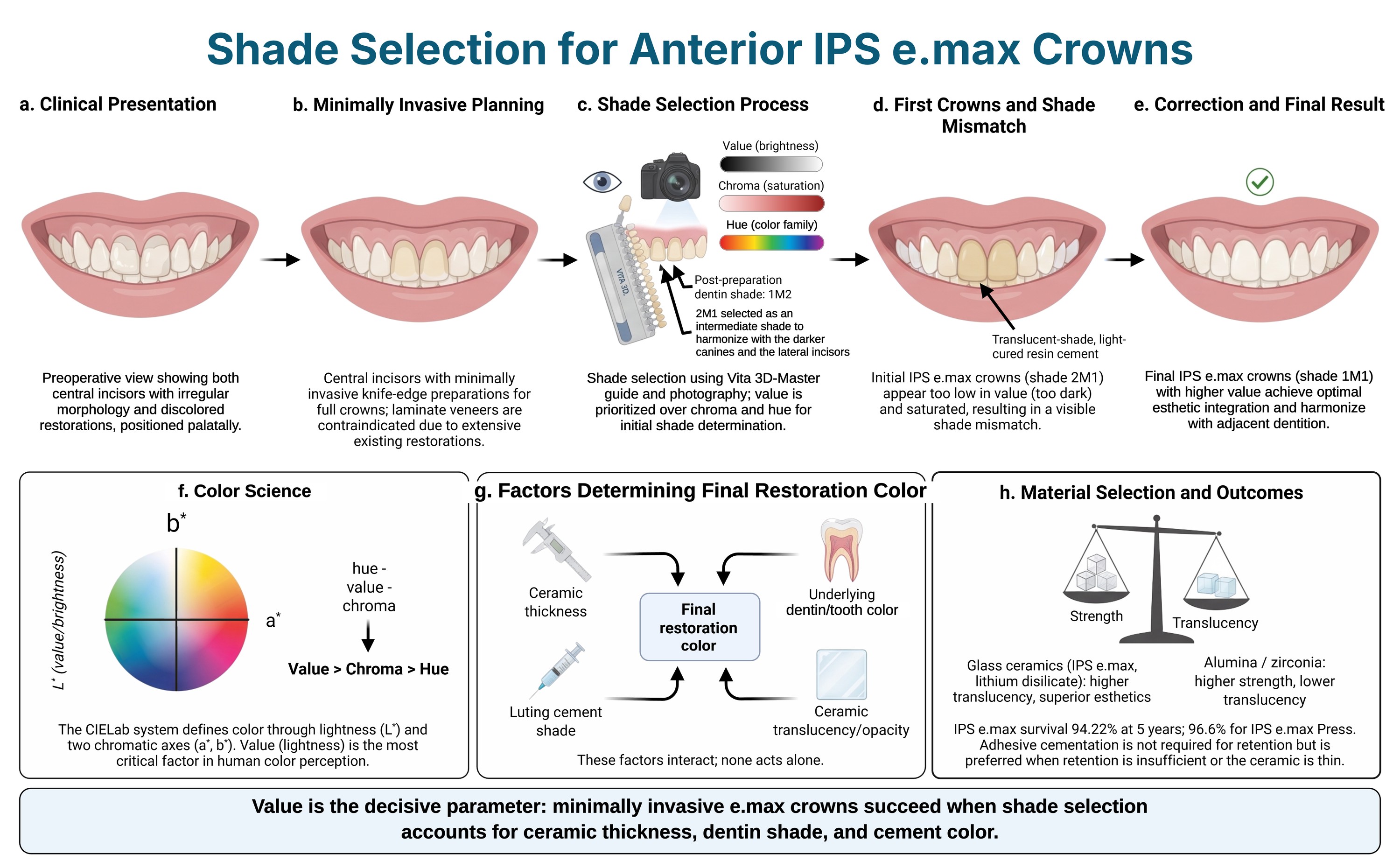
Introduction
Veneering of anterior teeth is a complex procedure (1). In general, patients have high esthetic expectations, and the final outcome largely depends on the skills of the dental technician (1). With increasing esthetic demands and expectations, the shade of the restoration has become the most critical factor in achieving ideal esthetics (2). The clinician must select the most appropriate shade for the patient and communicate it to the laboratory (2).
Accurate shade selection is a fundamental factor in achieving an acceptable esthetic smile (3). Tooth color occupies only a limited area within the visible light spectrum. Therefore, shade selection is particularly challenging for clinicians. The human eye can perceive light within the wavelength range of 380–760 nm. Color perception begins when light reaches the retina, where cone and rod cells are responsible for this function. There are three types of cone cells that perceive red, green, and blue light. Under conditions of insufficient light, rod cells become active, and color is not perceived. Color perception is a product of the brain and is influenced by various conditions. Consequently, the ability to perceive color varies among individuals. It has been observed that tooth color darkens with advancing age. When compared with other anterior teeth, the maxillary central incisors are lighter, whereas the maxillary canines are darker. The surface characteristics of teeth influence color perception by altering light reflection (3).
Color systems
Several systems have been developed over time to assess color characteristics (2). The Munsell Color System defines color in three dimensions: hue, value, and chroma (2, 4). Munsell considered hue to be the most easily understood variable and categorized it into five primary colors: purple, blue, green, yellow, and red. In the Vita classical shade guide, the primary colors are arranged into four groups: A, B, C, and D (2, 4). Value refers to the dimension that distinguishes between light and dark colors (2). On a scale from black to white, brightness increases from low to high. Brightness is defined as the amount of light reflected from an object; objects with high brightness appear less gray, whereas those with low brightness appear grayer. Chroma is the parameter that distinguishes a weak color from a strong one (2). It represents the saturation or pigment intensity of the hue (1). Chroma and brightness are inversely related (2). In the Vita classical shade guide, chroma is expressed numerically (2). Among the three fundamental components of color perception, value is considered the most important, followed by chroma and hue (5). The CIELab color system defines color on three axes (L*, a*, and b*) and is one of the most widely used systems (2). The L* coordinate represents the degree of lightness, with pure white corresponding to 100 and pure black to 0 (2, 4). The a* coordinate represents the red–green axis, where a positive a* indicates a tendency toward red and a negative a* indicates a tendency toward green (2). The b* coordinate represents the yellow–blue axis, where a positive b* indicates a tendency toward yellow and a negative b* indicates a tendency toward blue. The hue and chroma parameters of the Munsell color system do not directly correspond to the a* and b* coordinates of the CIELab system (2). The sequence of shade selection involves first determining value, followed by chroma, and finally hue (6).
Shade selection and shade matching
Shade selection methods can be classified into two categories: visual and instrumental (2). In visual shade determination, accuracy depends on variations in the light source, object, and observer. Factors such as the observer’s gender, age, cultural background, visual fatigue, and environmental conditions also influence the shade selection process. Tooth shade selection can be evaluated both subjectively and objectively. Visual shade selection using shade guides is subjective and has disadvantages, as factors such as the limited color range of the guide, observer-related influences, lack of standardization, and the absence of correspondence between the selected shade and the CIELab system significantly affect accuracy. In contrast, methods that employ devices for shade determination are considered objective. Instrumental techniques such as colorimeters, spectrophotometers, and image analysis methods are commonly used (2). Among these, red-green-blue (RGB) devices, such as ShadeScan, can generate a detailed color map of the tooth surface. Digital cameras provide rapid clinical support but are not sufficiently reliable as a stand-alone method. Spectrophotometers (e.g., SpectroShade, Vita Easyshade, ShadePilot) are regarded as the most accurate due to their light-independent and reproducible measurements. Colorimeters (e.g., ShadeVision, Shade NCC) use CIELab values for a more economical shade evaluation, although they are less precise than spectrophotometers (7). Shape, surface morphology, and value are the most important characteristics for achieving color harmony. Shade selection should be completed prior to tooth preparation, as dehydration may result in higher value readings. Value represents the most critical dimension in color determination (7). Shade guides used in dentistry include Vita Classical, Vitapan 3D-Master, and Chromascop, with the Vita 3D-Master being the most commonly used (2). In the Vita 3D-Master system, the sequence of shade selection follows the order of value, chroma, and hue. In addition to accurate shade determination, several other factors influence the final color of the restoration (2). The shade matching process should be carried out systematically to ensure accuracy, consistency, and predictable outcomes, which are essential in esthetic dentistry (6). Ceramic, the most commonly used restorative material, influences the final color of the restoration through its chemical composition, crystalline particle size, fluorescence, opacity, and translucency (2). In addition, the underlying tooth structure, ceramic thickness, and shade of the luting cement further affect the definitive color outcome (1). Try-in pastes, auxiliary materials that mimic the resin cement, can be used to preview how these factors affect the final color and to verify the color match before the restoration is cemented (8).
Restoration type selection
Although the aim of a minimally invasive approach is to perform restorations at the enamel level, in clinical situations such as tooth malposition, wear, or fractures, it may not be possible to remain within the enamel, and greater removal of the tooth structure may be required (9). However, a full crown is not always necessary in such cases. Translucent partial ceramic crowns can be considered an appropriate treatment option when restoration of both dentin and enamel is required and no modification of the existing occlusion or change in tooth shade is needed. In general, the reasons for using all-ceramic crowns in anterior teeth include the presence of previous restorations, situations where occlusion needs to be altered, or extensive proximal caries. In such cases, it may be challenging for the clinician to decide between selecting an opaque or translucent material. In cases of insufficient anterior guidance, the presence of parafunctional habits, or when a greater amount of tooth structure needs to be removed, the selection of materials with higher mechanical strength is recommended (9). Metal-ceramic restorations are recommended for patients with parafunctional habits (10). However, when a metal-free option is preferred, mechanically strong alumina-based ceramics or zirconia crowns can be considered (10). When the prepared tooth is of normal color, translucent materials can be preferred; they minimize tooth tissue loss, allow supragingival or equigingival margins, and provide an ideal bond because the ceramic is etchable (9). Fractured teeth, particularly in the anterior region, may not provide sufficient resistance and retention form for conventionally cemented restorations. For full crown restorations with dentin of acceptable color, preparation of an approximately 1 mm chamfer finish line and placement of the margin at either a supragingival or equigingival level are recommended. Mechanical requirements may vary depending on the presence of anterior guidance; accordingly, either translucent or opaque ceramics may be preferred. Opaque ceramics are more prominent when greater mechanical strength is required, and either adhesive resin cements or conventional cements may be used for cementation (9). Glass-based ceramics with low mechanical properties can achieve acceptable durability when luted with resin cements. In contrast, due to their superior mechanical properties, alumina- or zirconia-containing ceramics can be cemented using both conventional and resin cements (1).
All-ceramic restorations
All-ceramic materials are characterized by high biocompatibility and superior optical properties (11). With the advancement of technology, high-strength core materials used in all-ceramic restorations, such as alumina, zirconia, zirconia-reinforced alumina, magnesium-alumina spinel, and lithium disilicate, have been developed and are currently applied in clinical practice (9). Dental technicians layer ceramics on these core materials to provide restorations with their final esthetic appearance. All-ceramic systems can be classified into two main groups: translucent ceramics (conventional sintered feldspathic porcelain, pressable ceramics, and CAD/CAM ceramics) and opaque ceramics (alumina, zirconia, and lithium disilicate), which are used as high-strength core materials (9). In the presence of a full crown indication, the clinician should determine the ceramic material to be used (1). These materials are generally classified into two principal categories: acid-etchable glass ceramics with relatively low fracture resistance (e.g., IPS Empress, IPS e.max, and conventional feldspathic porcelain) and non-etchable ceramics with superior fracture resistance (e.g., alumina- or zirconia-based ceramics). In general, ceramics containing alumina or zirconia exhibit superior mechanical properties, whereas glass-based ceramics demonstrate enhanced optical characteristics (1). IPS e.max has a broad range of clinical applications. Owing to its glass-ceramic structure combined with lithium disilicate, it offers advantages such as not requiring adhesive cementation for retention, providing high esthetics, and exhibiting high fracture resistance (11). Both systems can be applied appropriately when the correct indication is established (1). The correct choice between them depends on the shade of the supporting tooth structure and the type of cementation system used. Glass-based ceramics exhibit higher translucency than zirconia- and alumina-containing ceramics. To effectively mask the underlying discoloration in the supporting tooth structure, approximately 2 mm of porcelain thickness is required. Therefore, in cases where discoloration of the underlying dentin is present, the use of glass-based ceramics is not considered suitable (1). In summary, translucent ceramics provide superior esthetics but lower mechanical strength, whereas opaque ceramics offer higher strength but reduced esthetic qualities (9). Therefore, the selection of all-ceramic materials should be made to achieve the most appropriate balance for each clinical case. In all-ceramic restorations, factors such as the color of the prepared tooth structure, indication, material type, translucency, and thickness influence the esthetic outcome (9). In addition, the luting cement has an influence on the final color of the restoration and should be carefully considered (2, 12). Dental ceramics are ranked from the most translucent to the most opaque as follows: IPS Empress 0.5 mm, In-Ceram Spinell 0.5 mm, IPS e.max 0.5 mm, IPS Empress 0.8 mm, Procera AllCeram 0.5 mm, IPS e.max 0.8 mm, In-Ceram Alumina 0.5 mm, and In-Ceram Zirconia 1 mm (10). The thickness of the material directly affects translucency; as the ceramic thickness increases, the brightness decreases, whereas the appearance of red and yellow tones becomes more pronounced. In anterior single-tooth restorations, the selection of the material depends on multiple esthetic factors. Among these factors, the translucency of the tooth, brightness, and the color of the underlying dentin tissue play an important role. The translucency and opacity of the adjacent tooth should be carefully evaluated. In cases where high translucency is required, glass-based ceramics provide a more similar esthetic outcome. In cases where the adjacent tooth is opaque, zirconia is recommended as the substructure material due to its ability to block light transmission and provide a uniform tone. The thickness of the zirconia substructure is less critical than that of glass-based ceramics. This is because the minimum cervical thickness required for glassy ceramics ranges from 0.8 to 1.5 mm, while for leucite, lithium disilicate, alumina-based ceramics, and zirconia, this value can be approximately 0.5 mm. If the tooth exhibits insufficient retention, resin-bonded cementation should be performed. Lithium disilicate-based ceramics are the first choice for achieving optimal bonding (10).
Case Presentation
A 32-year-old woman with no reported systemic disease presented with persistent dissatisfaction with the color and appearance of her maxillary central incisors. She reported a traumatic fracture of these teeth approximately 14 years earlier, followed by repeated composite resin restorations; the most recent restorations had been placed at another clinic 2 months before presentation. According to the patient, that clinic had recommended preparing all four maxillary incisors to achieve an acceptable esthetic match. Clinical examination revealed a deep bite and pronounced lingual inclination of the maxillary central incisors, which were positioned posterior to the smile line, exhibited morphological irregularities, and contained extensive composite resin restorations (Fig. 1). Both teeth were vital, and the periodontal tissues were clinically healthy, with no bleeding. Radiographic evaluation revealed no periapical radiolucency or other periapical pathology associated with the maxillary central incisors.
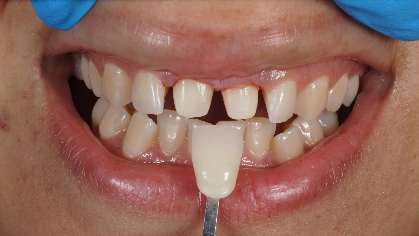
It was explained to the patient that the esthetic objectives could be achieved with a more conservative approach, and prosthetic treatment was planned only for the maxillary central incisors. Owing to the volume of the existing resin restorations, laminate veneers were not considered appropriate, and IPS e.max (Ivoclar, Schaan, Liechtenstein) all-ceramic crowns were selected to preserve hard tissue and achieve natural tooth-like esthetics. In view of the pronounced lingual inclination, buccal reduction was kept to a minimum, and a knife-edge finish line was selected to conserve cervical dentin and preserve the remaining sound tooth structure. Because of the translucency of IPS e.max, color was evaluated not only on the adjacent teeth but also on the prepared dentin surface. Shade selection was performed using the Vita 3D-Master shade guide (VITA Zahnfabrik, Bad Säckingen, Germany) and clinical photographs. The dentin shade of the prepared teeth was 1M2 (Fig. 2). Because the canines were darker than the lateral incisors, shade 2M1 was chosen to harmonize with both.
The crowns were fabricated from IPS e.max Press HT A1 ingots and veneered with IPS e.max Ceram using Dentin A1, Transpa blue, Transpa Incisal 1, and Opal Effect 2; the final total ceramic thickness was 1.5 mm. Before cementation, the shade match was evaluated with a try-in paste, and the patient approved the appearance of the crowns. The tooth surfaces were etched with phosphoric acid and the internal surfaces of the crowns with hydrofluoric acid. G-Multi PRIMER (GC Corporation, Tokyo, Japan) was applied to the internal ceramic surfaces, and G-Premio BOND (GC Corporation) was then applied to both the tooth surfaces and the internal surfaces of the crowns. The crowns were cemented with the Translucent shade of G-CEM Veneer light-cured resin cement (GC Corporation), which was selected because of the limited ceramic thickness and the high-translucency framework.
After cementation, both the patient and the clinician perceived the crowns as slightly darker than desired. Because the appointment took place in the evening, this appearance was initially attributed to the lighting conditions; however, the patient subsequently reported that the crowns also appeared too dark in daylight, and the restorations were considered unsuccessful in terms of color matching with the adjacent teeth (Fig. 3). Replacement crowns in shade 1M1, with a higher value, were requested using the same impressions and were fabricated with the same materials, except that Opal Effect 1 was used instead of Opal Effect 2. Approximately one week later, the initial crowns were removed, and the replacement crowns were cemented using the same surface-treatment and bonding protocol but with the Bleach shade of G-CEM Veneer. Because the concern prompting replacement was the final shade, the modifications were limited to the optical characteristics of the ceramic and the shade of the resin cement. The patient expressed satisfaction with the appearance immediately after the second cementation.
Follow-up examinations were performed 1 week, 1 month, and 6 months after the final cementation, and the esthetic outcome was assessed by visual comparison with a Vita shade guide, review of clinical photographs, and the patient’s subjective assessment. At 1 week, the shade match was considered satisfactory, and at the 1- and 6-month examinations, the restorations remained clinically satisfactory and integrated with the adjacent teeth in terms of color, form, and surface characteristics (Fig. 4). Written informed consent was obtained from the patient for the publication of this case report and the accompanying clinical photographs.



Discussion
The use of instrumental methods in shade selection has been found to be more reliable than the use of visual methods alone, and it has been recommended that both methods be used together whenever possible (13). Instrumental methods include various devices, such as colorimeters, spectrophotometers, intraoral scanners, smartphones, and digital cameras. Digital photography allows for a clear visualization of both the details of the tooth before treatment and the condition after treatment (13). A clinical study published in 2020 compared three shade determination methods: visual selection with the Vita 3D-Master guide, spectrophotometry with Vita Easyshade, and digital photography (2). Patient satisfaction with the all-ceramic restorations was 100% in the digital photography and Vita 3D-Master groups and 56% in the spectrophotometry group. Digital photography was more effective than the traditional visual method in terms of shade selection and surface characterization. It has also been shown that spectrophotometry cannot adequately record various tooth surfaces, although accurate results were obtained for monocolored teeth (2). A gray reference card, cross-polarizing filter, and light-correcting device for smartphones help standardize the light source during shade selection (13). Therefore, digital shade selection methods are considered more reliable than visual methods and allow the fabrication of ceramic restorations with more acceptable color differences (13). In a study measuring the precision and accuracy of four shade measurement devices, two spectrophotometers (SpectroShade and Vita Easyshade) and two digital cameras (ShadeVision and ShadeScan) were compared, with Vita Easyshade demonstrating the highest accuracy (2). In another study conducted in 2008, three spectrophotometers (Vita Easyshade Compact, Vita Easyshade, and ShadePilot) and one digital camera (ShadeVision) were evaluated, and the Vita Easyshade Compact device was found to have the best repeatability (2). Paravina reported that the Linearguide 3D-Master provided more accurate and user-friendly shade selection than other guides (14). Brandt et al. retrospectively evaluated 1,058 IPS e.max crowns and fixed partial dentures delivered in a private practice within three years and followed for up to 66 months (11). The most frequently fabricated restoration was IPS e.max Press, with 861 cases. In both recommended and non-recommended cases, including those with patient-related risk factors, IPS e.max restorations have shown high survival rates. The 5-year cumulative survival rate was 94.22%. Uses not recommended by the manufacturer were deliberately included, so that patient-related risk factors, such as poor oral hygiene and periodontal problems, could influence the outcome (11). In a similar study conducted by Yang et al., the 5-year cumulative survival rate of 6,855 IPS e.max Press restorations was reported to be 96.6% (15). In 2017, Rauch et al. reported a 6-year failure-free rate of 87.6% for lithium disilicate single crowns (16). In the literature, the clinical performance of glass-ceramic crowns has generally been reported as successful. Gemalmaz and Ergin followed 37 IPS Empress crowns for about two years and found that almost all restorations remained intact, with only one fracture recorded. They also observed that restorations with subgingival finish lines were more often associated with bleeding on probing than those with supragingival margins. This finding points to the importance of placing margins at or above the gingival level whenever possible to minimize biological complications (17). Dozić et al. examined the effects of varying thickness ratios of opaque and translucent porcelain layers in layered ceramic restorations. Their findings demonstrated that even minor changes in the OP/TP thickness ratio may result in perceptible color differences. They also reported that a 1-mm opaque layer could not completely mask the underlying background, whereas a 0.70-mm thickness of the tested core material was sufficient to eliminate this influence. These results underline the critical role of both layer thickness and background shade in achieving predictable esthetic outcomes (18). According to the literature, there is no definite conclusion regarding whether the shade of the resin cement affects the final color. Some studies have suggested that it has an effect, while others have reported no influence. Vichi et al. found that substrate-related color differences were not clinically relevant at an IPS Empress ceramic thickness of 2 mm, whereas at a ceramic thickness of 1 mm, color differences caused by the substrate and the cement were visually perceptible (19). Turgut and Bağış investigated the effect of different resin cement types and shades and of ceramic thickness on the final color of laminate veneers (12). They found that the color of the resin cement affected the final color of the restoration and that color differences were observed between resin cements with the same color label. They reported that the effect of the resin cement on the final color decreased as the ceramic thickness increased (12). In the study by Alghazali and colleagues, the influence of try-in pastes and resin cements on the final color of porcelain veneers was investigated (8). The researchers compared the outcomes of different shades of try-in pastes with those of resin cements of the same shade both before and after polymerization. Their findings showed that while try-in pastes are useful for anticipating the color of restorations, they do not always correspond precisely with the final appearance obtained after resin cement curing. Therefore, the evaluation of restoration color should not rely solely on try-in pastes but also include assessment with the resin cement itself (8).
Previous studies have pointed out that visual shade selection alone is limited and that combining it with instrumental techniques provides more consistent and reliable results (20). Several factors influence the accurate selection of shade (2). Shade guides have inherent limitations, and the use of different digital technologies can eliminate the subjectivity of shade selection (2). Compared with shade selection performed using a digital camera or smartphone, the visual shade selection method was found to fall below the acceptable threshold in terms of color matching between the ceramic crown and the adjacent tooth (13). The thickness of the restoration material and the color of the cement in all-ceramic restorations, together with properties such as the chemical composition of the ceramic, crystalline particle size, opalescence, fluorescence, and translucency, play a significant role in the formation of color (2). The choice of ceramic material for anterior restorations depends on several factors, including the required optical properties, amount of remaining tooth structure, margin placement, mechanical durability, and type of cementation (9). Translucent systems have been suggested as the closest answer to the question of which material is the most suitable in every case, with feldspathic and pressed ceramics highlighted for their strong bond to tooth structure (9). In this case, the shade initially considered clinically appropriate appeared darker than expected after cementation, suggesting the influence of factors such as material thickness, color of the underlying structure, and luting cement used (2). This highlights the need to support shade selection with visual shade guides, digital shade analyses, clinical try-ins, and, when required, try-in pastes (13). This case highlights that esthetic success in the anterior region depends not only on the restorative material itself but also on the proper management of the color–material–cement interaction (1, 2, 10). IPS e.max ceramics offer a reliable solution in the anterior region due to their translucency, color stability, and biocompatibility (9, 10). The careful implementation of shade selection protocols and close collaboration between the clinician and the laboratory are crucial for success (2). This clinical situation highlights the decisive role of the value (lightness) parameter in esthetic restorations (5, 7). For the long-term success of restorations, it is essential to consider and evaluate risk factors such as bruxism, poor oral hygiene, and periodontal problems (11).
Conclusion
In this case, where only the two maxillary central incisors were restored with IPS e.max crowns, a minimally invasive approach achieved esthetic harmony with the natural teeth despite the initial shade mismatch. The mismatch showed that shade selection in all-ceramic restorations is a multidimensional process that relies heavily on clinical experience and on the management of the color–material–cement interaction. Natural and satisfactory outcomes can be achieved even with restorations involving only two teeth; however, careful shade selection, with particular attention to value, and close collaboration between the clinician and the laboratory are essential.
Declarations
Acknowledgments: This case report was presented as a full-text paper at the 28th Turkish Dental Association International Dental Congress, Diyarbakır, Türkiye, September 18–21, 2025.
Informed Consent: Written informed consent was obtained from the patient for the publication of this case report and the accompanying clinical photographs.
Peer-review: Externally peer-reviewed.
Author Contributions: Conception – E.G.A.; Design – E.G.A., N.Ö.; Supervision – N.Ö.; Materials – N.Ö., E.G.A.; Data Collection and/or Processing – E.G.A.; Analysis and/or Interpretation – E.G.A., N.Ö.; Literature Review – N.Ö.; Writer – E.G.A.; Critical Review – E.G.A., N.Ö.
Declaration of Generative AI and AI-Assisted Technologies: During the preparation of this work, FigureLabs was used to create the graphical abstract, and the AI figure-generation feature in BioRender was used for subsequent text edits to the figure. The author(s) then reviewed, edited, and verified all visual and scientific content for accuracy and take(s) full responsibility for the final published material.
Conflict of Interest: No conflict of interest was declared by the authors.
Funding: The authors declared that this study has received no financial support.
Data Availability Statement: Data sharing is not applicable to this article as no datasets were generated or analyzed during the current study. The original patient records are not publicly available to protect patient confidentiality but are available from the corresponding author upon reasonable request.
References
- Mizrahi B. The anterior all-ceramic crown: a rationale for the choice of ceramic and cement. Br Dent J. 2008;205(5):251–255. https://doi.org/10.1038/sj.bdj.2008.735
- Yılmaz Ö, Dikicier S, Atay A. Tam seramik restorasyonlarda renk seçim yöntemlerinin ve etkileyen faktörlerin değerlendirilmesi: Literatür derlemesi. Dent & Med J - R. 2022;4(2):156–170.
- Önal B, Recen D, Türkün LŞ. Restoratif diş hekimliğinde renk seçimi. Turkiye Klinikleri J Restor Dent-Special Topics. 2015;1(3):21–27.
- Köroğlu A, Makhloota M, Turhan Bal B. A review of color matching in dentistry. Med Records. 2021;3(1):44–49. https://doi.org/10.37990/medr.818367
- Shaalan OO, Hashem BB, Khairy MA. Evaluation of shade matching of monochromatic versus polychromatic layering techniques in restoration of fractured incisal angle of maxillary incisors: a randomized controlled trial. J Int Oral Health. 2023;15(1):43–51. https://doi.org/10.4103/jioh.jioh_176_22
- Alnusayri MO, Sghaireen MG, Mathew M, Alzarea B, Bandela V. Shade selection in esthetic dentistry: a review. Cureus. 2022;14(3):e23331. https://doi.org/10.7759/cureus.23331
- Bhat V, Prasad DK, Sood S, Bhat A. Role of colors in prosthodontics: application of color science in restorative dentistry. Indian J Dent Res. 2011;22(6):804–809. https://doi.org/10.4103/0970-9290.94675
- Alghazali N, Moaleem M, Alamri S, Aldosari AA, Preston A, Smith P, et al. The effect of try-in paste and resin cement shade on colour properties of dental veneers. Eur J Prosthodont Restor Dent. 2018;26(3):144–151. https://doi.org/10.1922/EJPRD_01768Alghazali08
- Spear F, Holloway J. Which all-ceramic system is optimal for anterior esthetics?. J Am Dent Assoc. 2008;139(suppl):19S–24S. https://doi.org/10.14219/jada.archive.2008.0358
- Zaghba A, Soualhi H. Anterior ceramic crown: what ceramic for which situation. Int J Adv Res. 2019;7(12):1017–1022. https://doi.org/10.21474/IJAR01/10247
- Brandt S, Winter A, Lauer HC, Kollmar F, Portscher-Kim SJ, Romanos GE. IPS e.max for all-ceramic restorations: clinical survival and success rates of full-coverage crowns and fixed partial dentures. Materials (Basel). 2019;12(3):462. https://doi.org/10.3390/ma12030462
- Turgut S, Bagis B. Effect of resin cement and ceramic thickness on final color of laminate veneers: an in vitro study. J Prosthet Dent. 2013;109(3):179–186. https://doi.org/10.1016/S0022-3913(13)60039-6
- Jorquera GJ, Atria PJ, Galán M, Feureisen J, Imbarak M, Kernitsky J, et al. A comparison of ceramic crown color difference between different shade selection methods: visual, digital camera, and smartphone. J Prosthet Dent. 2022;128(4):784–792. https://doi.org/10.1016/j.prosdent.2020.07.029
- Paravina RD. Performance assessment of dental shade guides. J Dent. 2009;37(suppl 1):e15–e20. https://doi.org/10.1016/j.jdent.2009.02.005
- Yang Y, Yu J, Gao J, Guo J, Li L, Zhao Y, et al. Clinical outcomes of different types of tooth-supported bilayer lithium disilicate all-ceramic restorations after functioning up to 5 years: a retrospective study. J Dent. 2016;51:56–61. https://doi.org/10.1016/j.jdent.2016.05.013
- Rauch A, Reich S, Schierz O. Chair-side generated posterior monolithic lithium disilicate crowns: clinical survival after 6 years. Clin Oral Investig. 2017;21(6):2083–2089. https://doi.org/10.1007/s00784-016-1998-6
- Gemalmaz D, Ergin S. Clinical evaluation of all-ceramic crowns. J Prosthet Dent. 2002;87(2):189–196. https://doi.org/10.1067/mpr.2002.120653
- Dozić A, Kleverlaan CJ, Meegdes M, van der Zel J, Feilzer AJ. The influence of porcelain layer thickness on the final shade of ceramic restorations. J Prosthet Dent. 2003;90(6):563–570. https://doi.org/10.1016/s0022-3913(03)00517-1
- Vichi A, Ferrari M, Davidson CL. Influence of ceramic and cement thickness on the masking of various types of opaque posts. J Prosthet Dent. 2000;83(4):412–417. https://doi.org/10.1016/s0022-3913(00)70035-7
- Chu SJ, Trushkowsky RD, Paravina RD. Dental color matching instruments and systems: review of clinical and research aspects. J Dent. 2010;38(suppl 2):e2–e16. https://doi.org/10.1016/j.jdent.2010.07.001