Search
Search Results
##search.searchResults.foundPlural##
-
A Composite Laminate Veneer Technique for Diastema Closure: A Report of Ten Cases
Aim: Diastemas can be treated using periodontal, surgical, orthodontic, or prosthetic procedures. Composite laminate veneers can be applied to reduce or eliminate diastemas and represent a good aesthetic non-invasive alternative for these patients. This study presents the treatment results of 10 patients with diastemas between the anterior teeth of the maxilla and mandible with composite veneers. Methodology: Ten patients with diastemas between the anterior maxillary and mandibular teeth complaining of esthetic problems were treated at the Department of Operative Dentistry, University of Dicle. The periodontal health of the patients was acceptable and no caries were detected. Patients were informed of the treatment choices and composite resin veneers were selected as the better approach for each case. The presence of a diastema causes esthetic problems, especially in adolescents. Prosthetic and conservative methods are usually used to treat diastemas because these approaches are minimally invasive. The direct laminate technique has the advantages of low cost, treatment reversibility, and easy repair of the restoration intraorally. The direct composite laminate technique has become more effective because of improvements in adhesive dentistry. Conclusion: This study describes a direct composite laminate technique for the restoration of the anterior teeth of 10 patients with diastemas. These restorations were conservative and provide an inexpensive, one-visit aesthetic treatment for anterior teeth. How to cite this article: Bahşi E, Callea M, İnce B, Montanari M, Dallı M, Batteli F, Akdoğan M, Gabriella C, Toptancı İR, Gabriela P, Yavuz İ. A Composite Laminate Veneer Technique for Diastema Closure: A Report of Ten Cases. Int Dent Res 2012;2:67-74. Linguistic Revision: The English in this manuscript has been checked by at least two professional editors, both native speakers of English.Diastema closure, esthetic treatment, composite laminateEmrullah Bahşi, Michele Callea, Bayram İnce, Marco Montanari, Mehmet Dallı, Filippo Batteli, Mehmet Akdoğan, Clarich Gabriella, İsmet Rezani Toptancı, Piana Gabriela, İzzet Yavuz15.12.201267-74 -
Dentistry students after the COVID-19 outbreak—have views on distance education changed?
Aim: The aim of this study is to evaluate dentistry students for changes in anxiety levels between the initial outbreak of the COVID-19 pandemic and the point where COVID restrictions were reduced to a minimum and to compare their perspectives on distance education at both points in time. Methodology: The participants in this study were undergraduate students (n = 297) from the Faculty of Dentistry who volunteered to answer a questionnaire that asked them to detail the anxiety levels that they experienced during the COVID-19 period and during the months after COVID-related restrictions were lifted. The students were also asked for their opinions about the theoretical dentistry lessons delivered by distance education. The measurement values obtained were subjected to the independent sample t-test for comparisons between groups and to the one-way analysis of variance for comparisons of more than two groups. The Pearson correlation analysis was used to evaluate the relationship between anxiety levels. A value of p < 0.05 was considered statistically significant. Results: When asked whether technology-based education was beneficial, and whether theoretical education should be realized through distance education, men answered yes at a higher rate than women. ( p < 0.05). It was also observed that, compared to the early days of the COVID-19 pandemic, the anxiety scores of the participants were lower in the months after pandemic restrictions were reduced. Conclusion: According to the findings, it was observed that male students were more inclined to use the internet, to believe that technology-based education was beneficial, to prefer distance education over face-to-face education, and to be more motivated in distance education than face-to-face education. COVID-19 has impacted face-to-face education with a sudden transition to distance education, and these results support the importance of identifying the deficiencies in this education method and developing this system How to cite this article: Gönder HY, Fidan M, Demirel MG, Soybelli M, Alkurt S, Yüksel İB. Dentistry students after the COVID-19 outbreak—have views on distance education changed? Int Dent Res 2022;12(Suppl.1):34-43. https://doi.org/10.5577/intdentres.440 Linguistic Revision: The English in this manuscript has been checked by at least two professional editors, both native speakers of English.COVID-19, distance education, dental student, online education, surveyHakan Yasin Gönder, Muhammet Fidan, Mehmet Gökberkkaan Demirel, Mehmet Soybelli, Sinem Alkurt, İbrahim Burak Yüksel31.12.202234-43 -
Evaluation of parents’ awareness levels of traumatic dental injuries: The ToothSOS mobile application and “Save Your Tooth” posters
Aim: This survey study aimed to assess the awareness of parents of pediatric dental patients regarding the “ToothSOS” mobile application and the “Save Your Tooth” posters created by the International Association of Dental Traumatology. Methodology: This study included 303 participants who applied to Health Sciences University, Gülhane Faculty of Dentistry, Department of Pediatric Dentistry, between 02/01/2021 and 05/31/2021. The survey comprised visually supported questions to measure the participants’ demographic features, previous dental trauma experiences, awareness of mobile dental trauma applications, and knowledge of traumatic dental injuries (TDIs). Categorical variables were summarized as frequencies and percentages and were compared using the chi-square test ( p < 0.05). Results: The results showed that parents’ awareness levels regarding TDIs were insufficient. No significant difference was detected in the knowledge levels of parents of children with and without a history of dental trauma. However, significantly correct answers were obtained to the avulsion injury questions from parents with high education levels. Conclusion: Given the results of this study, it is recommended that dental health providers focus on TDI education for parents. How to cite this article: Bal C, Öztürk Z, Atak D, Yaşar M, Aksoy M. Evaluation of parents’ awareness levels of traumatic dental injuries: The ToothSOS mobile application and “Save Your Tooth” posters. Int Dent Res 2023;13(2):60-66. https://doi.org/10.5577/idr.2023.vol13.no2.3 Linguistic Revision: The English in this manuscript has been checked by at least two professional editors, both native speakers of English.ToothSOS, mobile app, dental trauma, pediatric dentistry, questionnaire, Dental Traumatology Treatment GuidelineCenkhan Bal, Zeynep Öztürk, Demet Atak, Merve Yaşar, Merve Aksoy31.08.202360-66 -
Performance of ChatGPT-5, Gemini 2.5 Flash, and DeepSeek-V3.2 large language models in response to questions about ozone in dentistry
Aim: The aim of this study was to compare and evaluate the accuracy and completeness of responses to open-ended questions about the use of ozone in dentistry that were addressed to three recent large language models (LLMs): ChatGPT-5, Gemini 2.5 Flash, and DeepSeek-V3.2. Methodology: A total of 25 open-ended questions on the subject of ozone in dentistry were asked to three different LLMs (ChatGPT-5, Gemini 2.5 Flash, and DeepSeek-V3.2). The chat history was deleted after each response, and a new session was started for each question. All questions were asked only once using the same computer IP address, on the same day, on the same fixed fiber Internet network. The questions were asked to the chatbots in English, and English responses were obtained and recorded. The responses were evaluated independently by two restorative dentistry specialists using two separate Likert scales for accuracy and completeness. In the statistical analyses, the Intraclass Correlation Coefficient (ICC) was calculated, and the Shapiro–Wilk, Kruskal–Wallis, Dunn–Bonferroni, and Spearman tests were applied. Results: The agreement between the evaluators was found to be high in respect of the accuracy points and varied according to the model for the completeness points. Significant differences were determined between the LLMs in the comparisons of the accuracy scores (p < 0.001). The median points of the ChatGPT-5 and DeepSeek-V3.2 models were determined to be statistically significantly higher than those of Gemini 2.5 Flash (p = 0.002 and p < 0.001, respectively). Significant differences were observed between the LLMs in terms of completeness scores (p = 0.004). In the subgroup analyses, the mean points of the ChatGPT-5 and DeepSeek-V3.2 models were determined to be statistically significantly higher than those of Gemini 2.5 Flash (p = 0.006 and p = 0.021, respectively). Conclusion: The responses to questions about the use of ozone in dentistry showed differences among the LLMs in terms of accuracy and completeness. ChatGPT-5 and DeepSeek-V3.2 demonstrated better performance than Gemini 2.5 Flash. Despite the high accuracy rates, it must not be forgotten that incorrect healthcare information can lead to serious outcomes. Therefore, it should be recommended that LLMs be used under human supervision in clinical applications and that future research focus on increasing the diagnostic reliability of artificial intelligence.Artificial intelligence, large language models, ChatGPT, Gemini, DeepSeek, ozone, dentistrySuzan Cangül, Özkan Adıgüzel, Makbule Taşyürek, Tuba Tunç, Hatice Ortaç31.12.2025165-174 -
Analyzing biomimetic dentistry YouTube videos' quality and content
Aim: This study evaluated the quality and content of YouTube videos about biomimetic dentistry. Methodology: A keyword search for "biomimetic dentistry" was conducted on YouTube. It used only publicly available Internet data from searching YouTube with the default filter “Biomimetic Dentistry” on April 3, 2023. Of the 200 examined videos, only 91 fit the inclusion criteria; those 91 videos underwent a content quality evaluation. The videos' viewing rates were determined after their demographic characteristics were assessed. The information quality index (VIQI) and the Global Quality Scale (GQS) were used to evaluate the video quality. For the statistical analysis, the Shapiro–Wilk, Kruskal–Wallis, Mann–Whitney U, Chi-square, and Fisher's exact tests were used. Results: Most of the videos (n = 59) were labeled "low content," and healthcare practitioners uploaded the majority (91.2%) of them to YouTube. The most discussed subject was advantages and disadvantages (87.9%), followed by the definition of biomimetic dentistry (76.9%). The high-content video group's GQS and VIQI scores were significantly higher than low-content group. There was a statistically significant positive correlation between the VIQI and GQS ( p < 0.05) ( p = 0,001) and a positive correlation between the overall content, total VIQI, and total GQS scores. Conclusion: The material in YouTube videos about biomimetic dentistry is insufficient for use as a patient information tool. Biomimetic dentistry-related YouTube videos' quality and content were typically poor. Health practitioners should be aware of the content of video-sharing platforms and take responsibility for enriching video content. How to cite this article: Erdem Hepşenoğlu Y, Değirmencioğlu D, Topbaş C. Analyzing biomimetic dentistry YouTube videos' quality and content. Int Dent Res 2023;13(S1):44-49. https://doi.org/10.5577/idr.2023.vol13.s1.7 Linguistic Revision: The English in this manuscript has been checked by at least two professional editors, both native speakers of English.YouTube, biomimetic dentistry, information quality index, global quality scaleYelda Erdem Hepşenoğlu, Duygu Değirmencioğlu, Celalettin Topbaş15.10.202344-49 -
Examination of stress distributions on Class V restorations using nanohybrid resin composite: A 3D finite element stress analysis
Aim: The aim of this study was to investigate and compare the distribution of stress zones in composite resins and dental tissues applied to different-sized Class V cavities in the cervical region of the tooth as a result of the forces acting on the tooth in a computer environment using the finite element method. Methodology: The extracted lower first molar tooth was scanned and recorded with a dental tomography device. Images were obtained as DICOM files, and these files were imported into the Mimics 12.00 program. In this program, a 3D image of the tooth was created and converted to Standard Tessellation Language (STL) files. The obtained STL files were transferred to the Geomagic Design X program, and Standard for the Exchange of Product Model Data (STP) files were created. The SolidWorks program was used to create different sizes of Class V cavity preparation, restorative material layers, adhesive material layers, and periodontal tissues. Finally, a finite element (FE) model was created in the ABAQUS program, and stress distributions were analyzed. Results: When the depth was kept constant and the width was increased, the amount of stress on the enamel tissue decreased, while the amount of stress on all other tissues and the restorative material increased. When the width was kept constant and the depth was increased, the amount of stress on the restorative material, adhesive layer, and cortical bone increased, while the amount of stress on the enamel layer, dentin layer, periodontal ligament, and trabecular bone decreased. Conclusion: Based on the values found in the study, the model that produced the least stress on the restorative material was the model with the least depth and width. However, the stress accumulation in the enamel tissue was higher in this model compared to the other models. As a result, the lifespan of restorative materials may be longer in cavities prepared more shallowly and narrowly during the preparation phase. How to cite this article: Harmankaya A, Gönder HY, Mohammadi R, Karaköy H, Tunçdemir MT, Yüksel İB, Göktolga EG. Examination of stress distributions on Class V restorations using nanohybrid resin composite: A 3D finite element stress analysis. Int Dent Res. 2024;14(S1):90-97. https://doi.org/10.5577/indentres.562Finite element analysis, nanohybrid, composite, Class V, Von Mises stressAbdulkadir Harmankaya, Hakan Yasin Gönder, Reza Mohammadi, Hilal Karaköy, Makbule Tuğba Tunçdemir, İbrahim Burak Yüksel, Emine Gülşah Göktolga31.12.202490-97 -
Evaluating the accuracy of ChatGPT and Gemini models in dental specialty exams: A comparative study of pediatric and restorative dentistry
Aim: The integration of large language models (LLMs) such as ChatGPT and Gemini into healthcare education presents novel opportunities for knowledge dissemination and assessment. Despite their growing prominence, empirical evidence regarding their reliability and domain-specific accuracy in dental specialty examinations remains limited. This study aimed to evaluate and compare the performance of four AI-driven LLMs—ChatGPT-3.5, ChatGPT-4o, Gemini 2.0 Flash, and Gemini 2.0 Advanced— by employing official past questions from the Turkish Dental Specialty Examination (DUS) in Pediatric Dentistry and Restorative Dentistry. Methodology: A total of 178 multiple-choice questions (90 in Restorative Dentistry and 88 in Pediatric Dentistry) were obtained from DUS examinations conducted between 2012 and 2017. All items were presented in Turkish to each model via standardized prompts in isolated sessions to prevent cross-interaction and memory bias. Response accuracy was determined by evaluating the correctness of the chosen answers. Comparative analyses between models, domains, and annual trends were conducted using non-parametric statistical tests, including the Mann–Whitney U and Friedman tests. Results: ChatGPT-4o achieved the highest overall accuracy across both Pediatric (90.88%) and Restorative Dentistry (93.02%), whereas ChatGPT-3.5 exhibited the lowest performance in Pediatric Dentistry (70.71%). Statistically significant inter-model differences were observed in Pediatric Dentistry (p = 0.002), but not in Restorative Dentistry (p = 0.254). Temporal analyses highlighted the consistent performance of ChatGPT-4o, while Gemini 2.0 Flash displayed marked fluctuations in accuracy across certain years and subdomains. Conclusion: Among the evaluated LLMs, ChatGPT-4o demonstrated superior and stable performance in addressing dental specialty exam questions, particularly in content requiring contextual comprehension. These findings underscore the potential of advanced AI tools to enhance dental education and examination readiness. Nonetheless, appropriate model selection should be tailored to the complexity of the task, domain specificity, and pedagogical objectives.Large language models, dental specialty examination, ChatGPT, Gemini, dental education, artificial intelligenceAyşenur Altuğ Yıldırım, Menekşe Alim28.06.2026e260673 -
Cleidocranial dysplasia. A molecular and clinical review
Cleidocranial dysplasia (CCD) is a rare autosomal dominant disorder characterized by skeletal and dental abnormalities primarily, short stature, aplasia or hypoplasia of clavicles, open fontanelles and supernumerary teeth. Heterozygous mutations of the runt related transcription factor 2 ( RUNX2 ) gene have been found in approximately 60-70% of cases leaving a large number of cases with no defined genetic cause which led us to delve into molecular mechanisms underlying CCD and thus to detect potential target genes to be explored in these patients. In this review we also highlight very broadly the phenotypic characteristics of previously reported patients with CCD. How to cite this article: Avendaño A, Cammarata-Scalisi F, Rizal MF, Budiardjo SB, Suharsini M, Fauziah E, Grande NM, Fortunato L, Plotino G, Yavuz I, Callea M. Cleidocranial dysplasia. A molecular and clinical review. Int Dent Res 2018;8(1):35-38. Linguistic Revision: The English in this manuscript has been checked by at least two professional editors, both native speakers of English.Cleidocranial dysplasia, RUNX2 gene, supernumerary teethAndrea Avendaño, Francisco Cammarata-Scalisi, Mochamad Fahlevi Rizal, Sarworini Bagio Budiardjo, Margaretha Suharsini, Eva Fauziah, Nicola Maria Grande, Leonzio Fortunato, Gianluca Plotino, İzzet Yavuz, Michele Callea30.04.201835-38 -
Impact of COVID-19 pandemic on pediatric dentistry treatment procedures
Aim: The SARS-CoV-2 (COVID-19) pandemic has forced pediatric dentistry to limit clinical procedures due to children’s role in spread of the virus and transmission routes in pediatric patients. In order to minimize contamination and cross-infection risk, dental examinations and treatments had been postponed and patient admission protocols rearranged. This study aimed to evaluate the effects of pandemic on the number of patients admitted and treated in the pediatric dentistry clinic of a university hospital in the one-year period following the March 2020 global pandemic declaration. Methodology: The study included pediatric patients aged 0-14 years admitted to a pediatric dentistry clinic of a faculty of dentistry between March 2019 and March 2021 in order to compare and make a retrospective evaluation of the one-year time intervals before and after the pandemic announcement in March 2020. Children were divided into the age groups 2-6 and 7-14. Evaluation parameters were chosen as examination, radiograph (panoramic and periapical), fissure sealing or topical fluoride application, compomer, composite filling, pulpotomy, root canal treatment, prefabricated crown, tooth extraction, and space maintainer. Results: The results of the study showed a statistically significant decrease in monthly average number of examinations after the declaration of the pandemic regardless of age and gender (p<0.001). In both age groups, there is a proportional decrease in pulpotomy, composite filling, fissure sealant or topical fluoride application, root canal treatment, compomer filling, prefabricated crown procedures during the pandemic compared to the pre-pandemic, while the number of filming procedures increased proportionally (p<0.001). Conclusion: Dental clinicians were advised to take on more radical treatment options during the pandemic. Further studies are needed to examine the effects of the statistical decrease in the number of procedures in the field of pediatric dentistry and treatment services on long-term community oral health. How to cite this article: Yüksel BN, Bezgin T. Impact of COVID-19 pandemic on pediatric dentistry treatment procedures. Int Dent Res 2021;11(3):140-8. https://doi.org/10.5577/intdentres.2021.vol11.no3.2 Linguistic Revision: The English in this manuscript has been checked by at least two professional editors, both native speakers of English.COVID-19 pandemic, dental treatment, pediatric dentistry, retrospective studyBurcu Nihan Yüksel, Tuğba Bezgin31.12.2021140-148 -
Ozone applications in dentistry: A review
Ozone (O 3 ) therapy has been applied in medicine and dentistry for many years and has become increasingly widespread. Ozone, composed of three oxygen atoms (O 3 ), is a strong antioxidant and a highly effective antimicrobial agent. Ozone gas has high oxidizing properties and exhibits a 1.5-fold higher antimicrobial effect than chloride. Its use has become popular in many fields of dentistry in recent years, owing to its various properties. It is categorized as one of the non-invasive, atraumatic, protective, and preventive treatment modalities that have been improved by modern dentistry, and its acceptability among patients has increased as a result. Ozone is an antimicrobial, analgesic, and biosynthetic agent that stimulates the immune system, enhances blood circulation capacity, and positively affects oxygen transport mechanisms. It influences both cellular and humoral immunity. All three forms of ozone (gaseous, oil, and solution) can be used in dentistry. It is indicated for the treatment of approximately 300 different pathological conditions. Ozone is used in endodontics (irrigation solution, intracanal medicament, abscess cases, disinfection), periodontology (gingivitis, periodontitis, periimplantitis, surgical injuries, prophylaxis), oral pathology (stomatitis, aphthous ulceration, candidiasis, herpes lesions), oral, dental, and jaw surgery (implantation, reimplantation, simple-to-complicated tooth extraction, homeostasis, wound healing, temporomandibular joint (TMJ) surgery, osteonecrosis cases), prosthetic dental treatment (disinfection of fixed and removable prostheses), restorative dental treatment (caries lesions, dentine hypersensitivity, cracked tooth, bleaching), orthodontics (remineralization and prophylaxis of white spot lesions), pedodontics (caries lesions, root canal treatment, positive psychological impact, short treatment duration), and sterilization of dental instruments and units. This review aimed to describe the usage fields and effects of ozone in dental applications, provide insights into clinical practices in other fields of dentistry, and introduce newly conducted in vitro and in vivo studies.Ozone, ozone therapy, dentistry, disinfectionMerve Doğan İlik, Özkan Adıgüzel, Myroslav Goncharuk-Khomyn31.12.2025222-236 -
Examination of stress distribution on Class I cavity restored using amalgam and bulk-fill composite: A 3D finite element stress analysis
Aim: The aim of this study was to examine the stress distribution on the tooth structure, restorative materials, and periodontal tissues prepared in a computer environment as a result of the forces acting on the mandibular premolar tooth surface, using the finite element stress analysis method. Methodology: The extracted mandibular premolar tooth was scanned with a dental tomography device and recorded. Images were obtained as digital imaging and communications in medicine (DICOM) files, which were transferred to the Mimics 12.00 program. This program was used to create a 3D image of the tooth, which was converted into a Standard Tessellation Language (STL) file. The resulting STL files were transferred to the Geomagic Design X program, some necessary adjustments, such as smoothing, were made, and Standard for the Exchange of Product Model Data (STP) files were created. The STP files were transferred to the SolidWorks program and Class I cavity preparation, restorative material layers, adhesive material layers, and periodontal tissues were created. Finally, the finite element (FE) model was created in the ABAQUS program, and the stress distributions were analyzed. Results: In the model restored with bulk-fill, compared to the model restored with amalgam, the amount of stress on dentin was found to be higher, while the amount of stress on cortical bone, trabecular bone, periodontal ligament, and enamel was found to be lower. In both models restored with bulk-fill composite and amalgam, the structure exposed to the most stress was the enamel layer, while the structure exposed to the least stress was the periodontal ligament. The amount of stress on the amalgam material was found to be higher than the amount of stress on the bulk-fill composite material. Conclusion: Amalgam and bulk-fill composite materials are frequently used today in posterior teeth with large cavities. As a result of our study, bulk-fill composite material caused less stress distribution than amalgam in all tissues except dentin tissue. How to cite this article: Karaköy H, Gönder HY, Mohammadi R, Harmankaya A, Tunçdemir MT, Yüksel İB, Göktolga EG. Examination of stress distribution on Class I cavity restored using amalgam and bulk-fill composite: A 3D finite element stress analysis. Int Dent Res. 2024;14(S1):98-105. https://doi.org/10.5577/indentres.570Finite element analysis, amalgam, bulk-fill, Class I, stress distribution, Von Mises stressHilal Karaköy, Hakan Yasin Gönder, Reza Mohammadi, Abdulkadir Harmankaya, Makbule Tuğba Tunçdemir, Emine Gülşah Göktolga11.10.202598-105 -
Evaluating the radiographic results of apexification treatment in children
Aim: This retrospective study aimed to evaluate the radiographic success of apexification treatments applied with different materials and techniques. Methodology: Periapical radiographs of 224 maxillary incisors in patients between 6 and 12 years of age who had undergone apexification treatment at the Dicle University, Faculty of Dentistry, Department of Pediatric Dentistry were retrospectively examined. Each apex was scored using the periapical index (PAI) on X-rays obtained at 12 months of follow-up after treatment, and the presence or absence of the apical barrier was evaluated. The Kruskal–Wallis H, Mann–Whitney U, and chi-square tests were used for the statistical analysis ( p < 0.05). Results: In the maxillary incisors, apexification was most commonly performed on the maxillary right central teeth (74.11%). The cause for this treatment was trauma in 97.77% of the patients. Enlargement in the periodontal space (95.98%), the presence of lesions (60.71%), loss of lamina dura (54.46%), and external resorption (43.75%) were observed. At 12 months of follow-up after treatment, apical closure (87.95%) was observed in all teeth with open apexes, and the ratio of patients with a PAI of < 2 was 89.35% for calcium hydroxide (CH) apexification and 92.73% for mineral trioxide aggregate (MTA) apexification. Conclusion: The radiographic success of apexification treatments with CH and MTA was found to be high in 224 immature upper incisors. How to cite this article: Akleyin E, Polat Y, Harman EB, Çelenk S. Evaluating the radiographic results of apexification treatment in children. Int Dent Res 2022;12(3):158-64. https://doi.org/10.5577/intdentres.2022.vol12.no3.8 Linguistic Revision: The English in this manuscript has been checked by at least two professional editors, both native speakers of English.Apexification, calcium hydroxide, mineral trioxide aggregate, periapical indexEbru Akleyin, Yelda Polat, Elif Burcu Harman, Sema Çelenk31.12.2022158-164 -
Retrospective evaluation of fissure sealants applied by dentistry students
Aim: The purpose of this study was to assess the adequacy and success in meeting protective expectations over time of resin-containing fissure sealants applied by dentistry students in our clinic. Methodology: Six-month controls were performed on 516 teeth of 85 patients aged 9–15 years. The 6-month controls of the resin-based pit and fissure sealants applied by 5th-grade dentistry students to patients presenting to the Necmettin Erbakan University Faculty of Dentistry Pedodontics Clinic between January 2022 and February 2022 were checked, and their survival was checked. The SPSS 26 statistical program was used in the analysis of the data. The statistical significance level in the study was determined as p < 0.05. A total of 85 patients and a total of 516 teeth, including premolars and molars, were included in the study. Results: A total of 128 teeth were processed in the right maxilla, and total retention was observed in 102 teeth. Right maxilla; When the left maxilla ( p = 0.001), left mandible ( p = 0.011), and right mandible ( p = 0.001) were compared in terms of total retention, a statistically significant difference was found. When the teeth were compared among themselves, the highest rate of loss, a total loss of 50%, was found in teeth 26 and 47. When the teeth were evaluated according to age, teeth numbered 14 ( p = 0.001) and 24 ( p = 0.001) were 9 years old, teeth 35 ( p = 0.001) and 45 ( p = 0.001) were 9–10 years old, and tooth number 44 (0.012). At the age of 10, the percentage of total loss was higher than in the other age groups, and a statistically significant difference was found. This suggests that the ideal isolation of the permanent premolars may not have been achieved because they do not fully erupt at the age of 9–10. Conclusion: When all the results were evaluated, it was seen that the retention of fissure sealants depended on multiple factors, and their indications should be carefully examined. How to cite this article: Abaklı İnci M, Özer H, Koç M. Retrospective evaluation of fissure sealants applied by dentistry students. Int Dent Res 2022;12(Suppl.1):59-64. https://doi.org/10.5577/intdentres.451 Linguistic Revision: The English in this manuscript has been checked by at least two professional editors, both native speakers of English.Pit and fissure sealant, caries, pediatric dentistry, preventive dentistryMerve Abaklı İnci, Hazal Özer, Merve Koç31.12.202259-64 -
Use of novel polyetheretherketone (PEEK) polymer in pediatric dentistry
The purpose of this study is to discuss the applications of PEEK in pediatric dentistry and the advantages of PEEK material in terms of periodontal health. In pediatric dentistry, there is a continuous search for biocompatible, esthetic, and durable materials to overcome the limitations of conventional options such as stainless steel and zirconia. Polyetheretherketone (PEEK), a high-performance semicrystalline polymer, has emerged as a novel alternative due to its superior mechanical properties, resistance to heat and chemicals, and an elastic modulus comparable to human bone. This review aims to evaluate the material characteristics of PEEK, its impact on periodontal health, and its clinical applications within pediatric dentistry. PEEK is amenable to digitalization and can be fabricated using CAD/CAM milling and 3D printing, ensuring precise fit and patient comfort. In pediatric orthodontics and space maintenance, PEEK serves as an esthetic, metal-free alternative, eliminating risks of nickel allergy while providing high strength and significantly reduced weight compared to traditional appliances. For restorative procedures, PEEK is utilized in crowns, endocrowns, and post-core systems. Its shock-absorbing capability allows for favorable stress distribution in dentin, reducing the risk of root fractures often associated with rigid materials like metal or glass fibers. Additionally, its low specific gravity and biocompatibility make it an effective material for obturators in treating cleft lip and palate defects. Regarding periodontal health, polished PEEK surfaces demonstrate low plaque affinity and acceptable surface roughness, supporting gingival health. PEEK offers distinct advantages in pediatric dentistry, particularly for patients requiring metal-free, lightweight, and shock-absorbing restorations. While it presents a promising alternative to traditional materials, achieving durable adhesion requires specific surface treatments, and further prospective clinical studies are warranted to validate its long-term performance.PEEK, pediatric dentistry, space maintainers, biocompatibility, CAD/CAM, periodontal health, dental applicationSemih Ercan Akgün, Şemsettin Yıldız, Samet Tekin31.08.202585-91 -
Treatment of hypomineralized maxillary central and canine teeth with resin infiltration technique: A case report
Aim: The resin infiltration technique is a minimally invasive approach that can be used to treat white spot lesions and arrest the progression of caries lesions. In this case report, it is aimed to eliminate the existing aesthetic problem by treating enamel hypomineralization in the maxillary central and canine teeth with the resin infiltration technique. Methodology: A 17-year-old male patient was referred to our clinic because of an aesthetic problem caused by a white spot lesion in his maxillary central and canine teeth. After clinical and radiological examination, his treatment options were evaluated, and we decided to treat it with the resin infiltration technique. After cleaning the surfaces of the teeth with a prophylaxis pad (Ultrapro Tx, Ultradent, South Jordan, UT, USA), isolation was achieved by applying a gingival barrier (Gingival Barrier, SDI, Bayswater, VIC, Australia). The adjacent teeth were protected using Teflon tape. A 15% hydrochloric acid gel (Icon Etch, DMG, Hamburg, Germany) was applied to the enamel surfaces for two minutes by mixing with a brush. After, the acid was removed using a water spray for 30 seconds. Ethanol (Icon Dry, DMG, Hamburg, Germany) was applied for 30 seconds and air dried. Then, a low-viscosity resin infiltrant (Icon Infiltrant; DMG, Hamburg, Germany) was applied to the tooth surfaces for three minutes by slight massage. After the application, the resin was light polymerized for 40 seconds (D-Light Pro, GC, Tokyo, Japan). The whole procedure was performed in such a way that there were two consecutive cycles. Results: Aesthetics improved after the procedure. The patient was followed up with control sessions. Conclusion: White spot lesions can be treated with a minimally invasive approach using the resin infiltration technique with appropriate indication. How to cite this article: Tüzel S, Çokkeçeci İT, Üçtaşlı MB, Arısu HD. Treatment of hypomineralized maxillary central and canine teeth with resin infiltration technique: A case report. Int Dent Res 2022;12(Suppl.1):124-7. https://doi.org/10.5577/intdentres.452 Linguistic Revision: The English in this manuscript has been checked by at least two professional editors, both native speakers of English.White spot lesion, hypomineralized tooth, aesthetic dentistry, minimally invasive, resin infiltration technique, Icon resin infiltrantSeden Tüzel, İlke Torbalı Çokkeçeci, Mine Betül Üçtaşlı, Hacer Deniz Arısu31.12.2022124-127 -
Shade selection for IPS e.max lithium disilicate crowns on maxillary central incisors: A case report and literature review
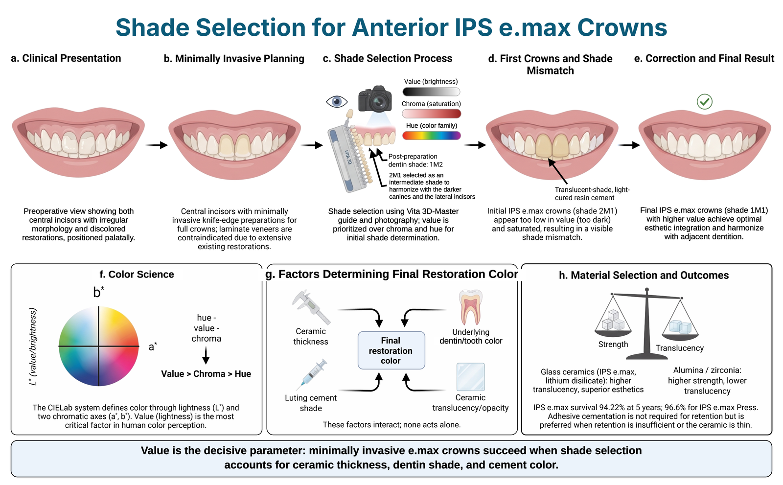
Shade selection is a critical step in the esthetic rehabilitation of anterior teeth, because the final appearance of an all-ceramic restoration depends on the selected shade, the ceramic material and its thickness, the color of the underlying tooth structure, and the luting cement. This case report and literature review describes the restoration of both maxillary central incisors with IPS e.max lithium disilicate crowns. A 32-year-old woman with a history of dental trauma and repeated composite restorations was dissatisfied with the appearance of her maxillary central incisors, which were lingually inclined, contained extensive composite resin restorations, and showed morphological irregularities. A conservative plan limited to these two teeth was adopted, and IPS e.max crowns were preferred over laminate veneers because of the volume of the existing restorations. After minimal buccal reduction with a knife-edge finish line, shade was selected with the Vita 3D-Master guide and clinical photographs, and the prepared dentin shade (1M2) was also recorded. The first crowns, fabricated in shade 2M1 and cemented with a translucent light-cured resin cement, appeared darker than the neighboring teeth. One week later, they were replaced with higher-value 1M1 crowns, which were cemented with a bleach-shade cement using the same bonding protocol. At the 6-month follow-up, the restorations blended with the neighboring dentition in color, form, and surface texture. The reviewed literature indicates that visual shade selection is subjective and is best combined with instrumental methods; that value is the most important dimension of color perception; and that ceramic thickness, substrate color, and cement shade can cause perceptible color differences in glass-ceramic restorations. Within the limitations of a single case, natural esthetic results can be achieved even when only two anterior teeth are restored, provided that visual and digital methods are combined, the color–material–cement interaction is managed, and clinician and technician communicate closely.Ceramics, color perception, crowns, dental esthetics, prosthesis coloring, resin cementsEkin Gökmen Akmansu, Nihal Özcan03.11.2026e260671 -
Evaluation of color stability after the coloring process in bulk-fill composites
Aim: Bulk-fill composites have been developed to overcome the disadvantages encountered in the incremental technique, such as the risk of contamination between layers, bonding problems, and gaps. Bulk-fill composites are frequently used in dentistry. The aim of this study was to evaluate the color stability of three different bulk-fill composites after a staining period with tea, coffee, red wine, and distilled water. Methodology: Three different flowable bulk-fill composites were used in the study (3M Filtek One, Dentsply Sirona SDR, Tokuyama Estellite Bulk-fill Flow). Composite disks with a diameter of 13 mm and a thickness of 1.5 mm were prepared for each subgroup using polytetrafluoroethylene molds. After polymerization, both surfaces of the samples were polished. The samples were incubated in distilled water at 37°C for 24 hours. Initial color measurements were taken using a spectrophotometer. The samples were then immersed in the coloring solutions for 72 hours. Color measurements were taken again, and ΔE 00 values were calculated using the CIEDE2000 formula. The data were statistically analyzed using three-way variance analysis (ANOVA) and Tukey HSD multiple comparison tests. Results: A significant difference in ΔE 00 values was observed between the three groups. The values in the SDR group were significantly higher than those of the 3M and Tokuyama groups. In the black tea group, the values were significantly lower than those of the red wine and coffee groups but significantly higher than those of the control group ( p <0.05). In the red wine group, the values were significantly lower than those of the coffee group but significantly higher than in the control group ( p <0.05). The values in the coffee group were significantly higher than those of the control group. Conclusion: Coloring solutions have an effect on the color stability of bulk-fill composites. Further studies and research should be done to improve bulk-fill composites. How to cite this article: Alkurt S, Gönder HY, Fidancıoğlu YD, Göktolga EG. Evaluation of color stability after the coloring process in bulk-fill composites. Int Dent Res. 2024;14(S1):50-54. https://doi.org/10.5577/indentres.560Composite, bulk-fill, color change, staining, CIEDE2000, flowable compositeSinem Alkurt, Hakan Yasin Gönder, Yasemin Derya Fidancıoğlu, Emine Gülşah Göktolga31.12.202450-54 -
PEEK in dentistry, properties and application areas
Aim: PEEK is a widely accepted material in medicine and dentistry. The aim of our study is to define and summarize the application areas of PEEK in dentistry. A retrospective literature research was conducted using the keywords ‘polyetheretherkethone’, ‘PEEK’, ‘dentistry’, ‘dental’,’ implant’, on the Pubmed and Google Scholar databases. The relevant articles were analyzed and summarized. In this review, the structure, properties and application areas of PEEK are explained. Conclusion: PEEK can be used in the biomedical field due to its excellent chemical and mechanical properties. Thanks to its bone-like elasticity modulus it can be used as an implant material, fixed and removable prostheses, obturators, pediatric dentistry, and orthodontics. In addition, PEEK is an alternative to metals due to its non-allergic and aesthetically acceptable properties. Nevertheless, improving PEEK's properties and increasing biocompatibility is a challenging process. It is expected that the use of PEEK in dentistry will increase due to the modifications applied. How to cite this article: Akay C, Ersöz MB. PEEK in dentistry, properties and application areas. Int Dent Res 2020;10(2):60-65. https://doi.org/10.5577/intdentres.2020.vol10.no2.6 Linguistic Revision: The English in this manuscript has been checked by at least two professional editors, both native speakers of English.PEEK, Polyetheretherkethone, Prosthodontics, İmplant, ThermoplasticsCanan Akay, Miraç Berke Ersöz31.08.202060-65 -
Shear bond strength of composite to demineralized enamel conditioned with resin infiltration
Aim: The aim of this in vitro study was to evaluate the influence of resin infiltration on bond-strength of composite resin to demineralized enamel. Methodology: Thirty bovine incisors were used in this study. Buccal enamel surfaces of bovine incisors were wet polished and then were divided into three groups: sound enamel; demineralized enamel; demineralized enamel infiltrated with a low-viscosity resin (ICON, DMG, Hamburg, Germany). After acid-etching with 37% phosphoric acid for 20 seconds, a two-step, total-etch adhesive (Single Bond 2, 3M ESPE, St. Paul, MN, USA) was applied using a microbrush for 20 seconds, followed by gentle air-drying for 5 seconds. The adhesive was light-cured for 10 seconds. Following the adhesive application, flowable composite resin (Filtek Supreme Flowable, 3M ESPE, St. Paul, MN, USA) was gently placed into a microtubule and was photopolymerized using an LED curing unit (Elipar Deep Cure; 3M ESPE, St. Paul, MN, USA). The microshear bond strength (µSBS) tests were performed using a microshear testing machine at a cross-head speed of 0.5 mm/min. One-way ANOVA and Bonferroni tests were used to analyze the data (5%). Results: Significant differences were found according to the ANOVA ( p < 0.05). Pair-wise comparison results of µSBS (mean ± SD) were: sound enamel (25.16 ± 2.3); demineralized enamel (17.93 ± 2.1); demineralized enamel infiltrated with a low-viscosity resin (28.51 ± 3.76). Conclusion: Resin infiltration applied to demineralized enamel before composite application increased the bond strength. No difference was found in the bond strength values obtained for sound enamel and resin infiltrated enamel. How to cite this article: Durukan SM, Gümüştaş B, Şişmanoğlu S. Shear bond strength of composite to demineralized enamel conditioned with resin infiltration. Int Dent Res 2022;12(Suppl.1):114-19. https://doi.org/10.5577/intdentres.441 Linguistic Revision: The English in this manuscript has been checked by at least two professional editors, both native speakers of English.Enamel, bond strength, resin infiltration, demineralization, remineralization, composite resinSebahat Melike Durukan, Burak Gümüştaş, Soner Şişmanoğlu31.12.2022114-119 -
Comparison of knowledge, attitude and practice toward oral health between pediatricians and family doctors
Aim: Pediatricians and family doctors are at the first step in the education of parents about oral health, as well as the prevention of oral diseases and the provision of oral health in children. The aim of this study is to evaluate the knowledge, attitudes and practices of pediatricians and family doctors about oral health in children. Methodology: A questionnaire consisting of 50 items was sent to pediatrician and family doctors via mail. Descriptive analysis and chi-square tests were done to analyze the data. Participation in the survey, with a total of 240 participants (58 pediatricians, 182 family doctors), was 100%. Results: It was found that pediatricians believe that they have more important role in the development of oral health and prevention of oral diseases and they are more interested in participating in the oral health program than are the family doctors. Conclusion: Pediatricians and family doctors should work with dentists to improve the quality of oral hygiene in all young children. How to cite this article: Gezgin O, Korkut E, Özer H, Şener Y. Comparison of knowledge, attitude and practice toward oral health between pediatricians and family doctors. Int Dent Res 2018;8(1):7-14. Linguistic Revision: The English in this manuscript has been checked by at least two professional editors, both native speakers of English.Children, family doctor, oral health, pediatricianOnur Gezgin, Emre Korkut, Hazal Özer, Yağmur Şener30.04.20187-14 -
Areas for use of PEEK material in dentistry
Polyetheretherketone (PEEK) material is a polycyclic, aromatic, thermoplastic polymer that is semi-crystalline and has a linear structure. PEEK has good mechanical and electrical properties such as resistance to high temperature and resistance to hydrolysis. In addition, because of the property of high biocompatibility, use of PEEK has increased in orthopedic and trauma cases. The most characteristic property of PEEK material is that it has a low elasticity modulus, close to that of bone. It has been suggested that stress-based problems could be reduced with this material due to the low elasticity modulus. In the light of this information, PEEK material could be considered as an alternative to conventional materials in the field of dentistry. How to cite this article: Tekin S, Cangül S, Adıgüzel Ö, Değer Y. Areas for use of PEEK material in dentistry. Int Dent Res 2018;8(2):84-92. Linguistic Revision: The English in this manuscript has been checked by at least two professional editors, both native speakers of English.Polyetheretherketone, PEEK, polymer, dental application, dental implantSamet Tekin, Suzan Cangül, Özkan Adıgüzel, Yalçın Değer27.08.201884-92 -
Evaluation of shear bond strength of amalgam restorations repaired with composite resin by different surface treatments
Aim: Repairing amalgam restorations with composite resins is a clinical practice with many advantages. An effective adhesion is achieved between amalgam and composite by applied different surface treatment on amalgam surface. In this study, the effect of adhesive systems applied on amalgam surface on composite-amalgam connection was investigated. Methodology: Standard cavities were opened in 70 acrylic molds that had completed their polymerization and amalgams were placed in these cavities. Seven groups were randomly assigned to different surface treatments on amalgam. In group 1 only roughening with diamond bur , in group 2 acid etch, in group 3 acid application after roughening with diamond bur, in group 4 roughening with diamond bur and silane, in group 5 acid etch and silane, in group 6 roughening with diamond bur and Alloy Primer and finally 7. acid etch and Alloy Primer was applied in the samples. After the application of clearfil universal bond to all groups, composite resins were placed and polymerized. Shear bond strengths were tested with a Universal testing machine. Obtained data were evaluated by one way ANOVA and Tukey B tests. Results: In the statistical evaluation of the groups, the highest shear bond strength between amalgam-composite was seen in the 3rd group where both roughening with diamond bur and acid were applied together, and the lowest shear bond strength was seen in the first group with only roughening with diamond bur. The differences between the groups were statistically significant (p <0.05). Conclusion: The roughening of the amalgam surface affects the shear bond strength between amalgam and composite resin. These results obtained in vitro conditions should be supported with clinical studies. How to cite this article: Cangül S, Adıgüzel Ö, Erpaçal B, Ünal M, Acartürk M, Satıcı Ö. Evaluation of shear bond strength of amalgam restorations repaired with composite resin by different surface treatments. Int Dent Res 2019;9(3):105-10. Linguistic Revision: The English in this manuscript has been checked by at least two professional editors, both native speakers of English.amalgam, composite, shear bond strengthSuzan Cangül, Özkan Adıgüzel, Begüm Erpaçal, Mehmet Ünal, Musa Acartürk, Ömer Satıcı31.12.2019105-110 -
Dyskeratosis congenita: A report of two cases
Dyskeratosis congenita (DC) is the first genetic syndrome identified among telomeropathies. Its classical phenotype is characterized by mucocutaneous abnormalities, atrophy and pigmentation of the skin, oral leukoplakia, and nail dystrophy. Bone marrow failure and susceptibility to malignancy are rare, and DC is mostly an X-linked recessive, serious, multisystemic disease. This study aims to increase dentists’ awareness of this genetic disease, accompanied by oral findings. Two cases of DC are reported in this paper, along with a short review of the literature. Oral findings of the disease include dental caries, gingival recession, short roots, gingival bleeding, severe mobility of the teeth, and alveolar bone loss. DC is clinically seen mostly in males aged 5 to 12. This case report examines dermatological and oral findings consistent with the disease of two brothers diagnosed with DC who applied to the Gülhane Health Sciences University, Faculty of Dentistry, Pediatric Dentistry Department for dental caries complaints. DC is a rare disease, and dentists should pay attention to its systemic and oral symptoms during their examinations. Despite a positive prognosis of this disease, it is important to take into account any unexpected changes in hematological values and mucocutaneous malignant alterations. How to cite this article: Aydın Varol E, Kılıç G, Bal C, Atay İ. Dyskeratosis congenita: A report of two cases. Int Dent Res 2023;13(3):133-7. https://doi.org/10.5577/idr2023.460 Linguistic Revision: The English in this manuscript has been checked by at least two professional editors, both native speakers of English.Dyskeratosis congenita, oral findings, oral health, leukoplakia, keratosisEzgi Aydın Varol, Gülşen Kılıç, Cenkhan Bal, İrem Atay31.12.2023133-137 -
Evaluation of liquid sorption and color stability of dental composites after exposure to common lactation teas
Aim: To assess the effects of common lactation teas on liquid sorption and color stability of three different dental composites. Methodology: A total of 60 samples (n = 5) were examined from three composite materials: Omnichroma (Tokuyama Dental Co., Tokyo, Japan), Estelite Posterior, (Tokuyama Dental Co., Tokyo, Japan), and Mosaic Universal (Ultradent production Inc., South Jordan, UT, USA). Samples measuring 2x6 mm were taken and immersed in three different teas—Humana (Humana, Bremen, Germany), Hipp, (Hipp, Pfaffenhofen, Germany), and Lactamil (Nutricia, Friedrichsdorf, Germany) —as well as artificial saliva. The measurements were recorded at baseline and on the 7th day. A sensitive analytical balance was used to measure liquid sorption, and a VITA Easy Shade device was used for color measurements. Absorption and Delta E values were calculated. The data were analyzed using MANOVA at a significance level of (p<0.05). Results: Composite materials, solutions, and their interactions had a statistically significant effect on the sorption and Delta E values (p<0.001). The relative liquid absorption values among the composites were recorded as follows: Tokuyama>Mosaic>Omnichroma. Humana had the highest and Hipp had the lowest mean value among the solutions (p<0.001). For Delta E, Mosaic had the highest mean value among the composites, and Lactamil had the highest mean value among the solutions (p<0.001). Conclusion: Lactation teas cause discoloration in dental composites. In vitro and in vivo studies on color changes in dental composites are needed due to the sorption of these fluids. How to cite this article: Yıldırım Bilmez Z, Şeker O, Köse HD, Aslan Gözüyeşil B.Evaluation of liquid sorption and color stability of dental composites after exposure to common lactation teas.Int Dent Res 2021;11(Suppl.1):228-33. https://doi.org/10.5577/intdentres.2021.vol11.suppl1.33 Linguistic Revision: The English in this manuscript has been checked by at least two professional editors, both native speakers of English.sorption, color change, composite, lactation teaZuhal Yıldırım Bilmez, Oya Şeker, Hazal Deniz Köse, Başak Gözüyeşil Aslan30.09.2021228-233 -
Microleakage of two different posterior composites and a high-viscosity glass-ionomer cement: An in vitro study
Aim: Using AutoCAD, we examined the microleakage of permanent molars in Class I restorations using a conventional posterior composite resin, a bulk-fill composite resin, and a high-viscosity glass-ionomer cement (HVGIC). Methodology: In total, 33 extracted noncarious third molars were used. Class I cavities were prepared. The teeth were randomly divided into three groups of 11 teeth each, as follows: Group A (Filtek Z550), Group B (Filtek Bulk-Fill Posterior Restorative), and Group C (EQUIA Fil). All specimens were stored for 24 h at 37 °C in distilled water. The teeth were thermocycled 1,000 times between 5 ± 2°C and 55 ± 2°C prior to immersion in 0.5% basic fuchsin for 24 h. Two mesiodistal cuts of each tooth were photographed under a stereomicroscope equipped with a digital camera. The dye-infiltrated surface area was measured. Results: Statistical evaluations were performed by one-way analysis of variance [ANOVA]. One-way ANOVA showed no significant difference between the three groups in microleakage (P = 0.07). However, the lowest microleakage ratio was seen in Group C (EQUIA Fil). Similar results were obtained with all tested materials, but the lowest microleakage rate was obtained with EQUIA Fil (Group C). Conclusion: In this study, the tested materials were not completely successful in eliminating microleakage, although the lowest microleakage rate was obtained with EQUIA Fil. How to cite this article: Çelenk S, Ataş O, Ayna B, Günay A. Microleakage of two different posterior composites and a high-viscosity glass-ionomer cement: An in vitro study. Int Dent Res 2022;12(1):34-8. https://doi.org/10.5577/intdentres.2022.vol12.no1.6 Linguistic Revision: The English in this manuscript has been checked by at least two professional editors, both native speakers of English.composite resin, high-viscosity glass-ionomer cement, microleakage, AutoCADSema Çelenk, Osman Ataş, Buket Ayna, Ayşe Günay30.08.202234-38 -
The use of micro-computed tomography in dental applications
Together with developments in digital imaging systems, there has been increasing use of micro-computed tomography (μCT) in many areas, primarily in dentistry. As this method allows 3-dimensional images to be taken providing safe and high quality results, without damaging the samples being examined, it is often preferred. Consequently, it has become possible to record rapid progression in endodontic studies in particular and to research and easily compare several techniques. Furthermore, this method can be used in tissue engineering, forming data for FEM analysis, the growth and development of craniofacial bones, imaging bone structure, and in the evaluation of implants and surrounding bone in root tip surgery, to determine mineral concentration in the teeth and to measure the thickness of enamel. The aim of this review was to focus on the areas of use of μCT in dentistry and state the advantages. Key Words : Micro-computed tomography, X-ray micro-CT, dental application, three-dimensional imaging How to cite this article: Erpaçal B, Adıgüzel Ö, Cangül S. The use of micro-computed tomography in dental applications. Int Dent Res 2019;9(2):78-91. Linguistic Revision: The English in this manuscript has been checked by at least two professional editors, both native speakers of English.Micro-computed tomography, X-ray micro-CT, dental application, three-dimensional imagingBegüm Erpaçal, Özkan Adıgüzel, Suzan Cangül31.08.201978-91 -
Evaluation of surface roughness of composites obtained by additive and subtractive manufacturing methods after sandblasting
Aim: The aim of this study was to observe the change in the surface roughness of composite samples produced by different production methods after sandblasting with aluminum oxide particles and to evaluate the effect of this on the composite material. Methodology: The study tested three subtractive method composites (GC Cerasmart [GC], Vita Enamic [VE] and Grandio Voco [GV]) and one additive method composite (3D Formlabs [3D]) were used. Each subtractive composite had 11 samples (14 x 14 x 2 mm) prepared with an Isomet low-speed saw, whereas the additive composite had 11 specimens produced using an SLA-based 3D printer (Form 2; Formlabs Inc). For roughness measurements and sandblasting, the samples were marked with an adhesive circular paper. Surface roughness was measured using a MarSurf SF1 profilometer, with three readings per sample. Sandblasting with aluminum oxide particles (50 µm) was performed using an Aquacare twin in-mouth sandblaster, and surface roughness measurements were repeated. Statistical analysis was performed using one-way analysis of variance (ANOVA) via SPSS 26, with significance set at p =0.05. Results: Among the composite samples, Vita Enamic (VE) had the highest average roughness value, and 3D Formlabs (3D) had the lowest. VE had a mean roughness value of 0.4575 µm, GC had a mean roughness value of 0.4627 µm, GV had a mean roughness value of 0.665 µm and D had a mean roughness value of 0.4575 µm. All groups were compared pairwise with each other. A significant difference was found between the GV-D, VE-D, GC-GV, GC-VE, and GC-D group pairings ( p <0.05). There was no significant difference between the GV-VE and GC-D group pairings ( p >0.05). Conclusion: Sandblasting with aluminum oxide was observed to increase the roughness of the materials, and the roughness varied depending on the nature of the material. Further studies are required to investigate the effects of roughness on the bonding. How to cite this article: Güntekin N, Alkurt S, Çiftçi A, Gönder HY, Büyükerkmen EB. Evaluation of surface roughness of composites obtained by additive and subtractive manufacturing methods after sandblasting. Int Dent Res. 2024;14(S1):60-64 https://doi.org/10.5577/indentres.559CAD-CAM, composite, surface roughness, wear, sandblasting, 3D printingNeslihan Güntekin, Sinem Alkurt, Aslı Çiftçi, Hakan Yasin Gönder, Emine Begüm Büyükerkmen31.12.202460-64 -
A micro-computed tomography evaluation of the change in volume of different bulk-fill composite materials caused by polymerization shrinkage
Aim: Composite resins contain different monomers and fillers that are directly affected by polymerization shrinkage. Therefore, an accurate and reliable method is needed to measure the volume changes. The aim of this study was to evaluate the amount of volume change associated with polymerization shrinkage in four different bulk-fill composite materials in class II restorations using a micro-CT device, which has high resolution and provides 3-dimensional images. Methodology: A total of 40 human 3rd molar teeth were used. First, standard class II cavities were opened on the mesial surfaces of all the teeth, and then the first micro-CT images were obtained. The same adhesive material was applied to all the teeth. The teeth were then separated into four groups, and a different bulk-fill composite was applied to each group; Filtek (FTK), X-tra Fil (XTF), Tetric Evo Ceram (TEC), and Filtek One (FLO) and the second micro-CT images were obtained. Then after polymerization of the materials, the final micro-CT images were taken, and analyses were made according to the scanning results. The Kruskal Wallis and Mann Whitney U-tests were used in the statistical evaluation of the data. Results: The volumetric gap formed after polymerization of the composite resins was not determined to be statistically significant (p>0.05). Conclusion: The volumetric difference (%) between the composite resin and the dental tissue following polymerization was seen to be greatest in XTF and least in FTK. It was concluded that the volumetric gap caused by the polymerization shrinkage of the tested materials may be due to the structure of the material. How to cite this article: Cangül S, Adıgüzel Ö, Sağmak S, Evran B. A micro-computed tomography evaluation of the change in volume of different bulk-fill composite materials caused by polymerization shrinkage. Int Dent Res 2021;11(2):75-82. https://doi.org/10.5577/intdentres.2021.vol11.no2.4 Linguistic Revision: The English in this manuscript has been checked by at least two professional editors, both native speakers of English.Micro-CT, bulk-fill composite, volumetric gap, polymerization shrinkageSuzan Cangül, Özkan Adıgüzel, Savaş Sağmak, Begüm Evran31.08.202175-82 -
Comparative evaluation of apical pressure generated by EDDY, GentleWave G4, and passive ultrasonic irrigation: A 3D-printed root canal model
Aim: This in vitro study compared the mean and peak apical pressures generated by EDDY, GentleWave G4, and passive ultrasonic irrigation (PUI) in a standardized 3D-printed root canal model. Methodology: A standardized straight root canal model (apical diameter 0.25 mm, 4% taper, 16 mm working length) was fabricated by resin-based stereolithographic printing. Apical pressure was measured with a validated setup comprising a piezoresistive pressure transducer (PXM409-350; Omega), a DAQ board (USB6008; National Instruments), and LabVIEW software. Distilled water served as the irrigating medium. Twenty measurements were performed per group (n=20, 60 total), with each recording 12,020 data points over 12 seconds. EDDY was activated with a TA-200 air scaler (power level 2, #25/.04 tip); GentleWave G4 was operated per the manufacturer's protocol; and PUI was performed with a Newtron P5 XS ultrasonic unit (power level 6, IrriSafe #20 tip, 2 mm short of the working length). Normality was confirmed by the Shapiro-Wilk test, and group comparisons were performed using Welch ANOVA with Games–Howell post hoc comparisons. The study followed PRILE 2021 reporting guidelines. Results: Significant differences were observed across all groups for both mean and peak apical pressures (p < 0.001). EDDY produced the highest mean (16.03 ± 4.04 mmHg) and peak (35.53 ± 2.84 mmHg) apical pressures, exceeding the safety threshold in every measurement. GentleWave G4 yielded a negative mean apical pressure (−15.60 ± 7.96 mmHg) and a mean peak of 8.00 ± 6.76 mmHg, remaining below the threshold throughout. PUI generated near-zero mean apical pressure (−0.07 ± 0.05 mmHg) and a mean peak of 2.27 ± 0.05 mmHg, consistently below the critical threshold. All pairwise comparisons were significant (p < 0.001), including GentleWave G4 vs. PUI for peak pressure (p = 0.003). Conclusion: GentleWave G4 and PUI produced apical pressures below the established safety threshold, whereas EDDY exceeded it in all measurements, indicating a higher risk of periapical irrigant extrusion. Further ex vivo and in vivo studies with anatomically complex geometries are warranted.Apical pressure, endodontic irrigation, GentleWave G4, irrigant extrusion, sonic activation, PUIHande Özyürek, Taha Özyürek, Olcay Özdemir, Ali Aminimofrad, Özkan Adıgüzel, Mustafa Gündoğar11.06.2026e260742 -
Evaluation of the dental anxiety levels of patients applying to the faculty of dentistry during the COVID-19 pandemic
Aim: The purpose of this study is to determine the levels of dental anxiety in patients who applied to the Necmettin Erbakan University Faculty of Dentistry during the COVID-19 outbreak and to evaluate the relationships of their anxiety levels to age, gender, education level, and the frequency of visits to dentists. Methodology: There were 320 participants, including 163 females (50.9%) and 157 males (49.1%), aged from 18 to 65 years or older, who had applied to the Necmettin Erbakan University Faculty of Dentistry. The Corah Dental Anxiety Scale (CDAS) was used to determine the levels of dental anxiety (non-anxiety, little anxiety, anxiety, over anxiety, and excessive anxiety) and the anxiety groups (low or high). Descriptive statistics were presented in percentages. Data were analyzed using t-tests and one-way ANOVA and chi-square tests, at a significance level of p = 0.05. Results: Among the participants, 38.1% were at the non-anxiety level, 44.1% had very little anxiety, 10.9% had anxiety, 5% had over anxiety, and 1.9% had excessive anxiety. With regard to the anxiety groups, 82.5% were in the low anxiety group, and 17.5% were in the high anxiety group. Age, education level, and frequency of visiting the dentist were not found to have any effects on dental anxiety (p > 0.05). However, the effect of gender with regard to dental anxiety was significant (p < 0.05), as females had higher dental anxiety levels than males. Conclusion: While the effect of gender was found to be important in terms of dental anxiety, age, educational status, and the frequency of visiting the dentist did not have significant effects on dental anxiety. Females had higher dental anxiety scores and levels than males. How to cite this article: Tunçdemir AR, Büyükerkmen EB, Savaş A. Evaluation of the dental anxiety levels of patients applying to the faculty of dentistry during the Covıd-19 pandemic. Int Dent Res 2021;11(Suppl.1):238-244. https://doi.org/10.5577/intdentres.2021.vol11.suppl1.35 Linguistic Revision: The English in this manuscript has been checked by at least two professional editors, both native speakers of English.dental anxiety, COVID-19, age, gender, education, dental visitAbdulhaluk Savaş, Emine Begüm Büyükerkmen, Ali Rıza Tunçdemir30.09.2021238-244 -
A Fibre-Reinforced Fixed Partial Denture on a Hemisectioned Tooth: A Case Report
In modern dentistry, fibre-reinforced fixed dental prostheses are considered a useful alternative to classical metal-ceramic restorations. This method allows a conservative approach for replacing missing teeth that overcomes some of the drawbacks of conventional prostheses. Our patient required extraction of tooth #46 because of an apical lesion of the mesial root, and underwent extraction by hemisection. After healing, using the superior properties of the combined fibre/composite, an adhesive bridge restoration was applied with support from the distal root of tooth #46 and teeth #45 and #47. How to cite this article: Agüloğlu S, Ayna E, Özdemir E. A Fibre-Reinforced fixed partial denture on a hemisectioned tooth: A Case Report. Int Dent Res 2011;1:38-41. Linguistic Revision: The English in this manuscript has been checked by at least two professional editors, both native speakers of English.Fibre-reinforced, adhesive bridge, hemisectionSüleyman AGÜLOĞLU, Emrah AYNA, Eylem ÖZDEMİR15.04.201138-41 -
The relationship between mandibular symphysis shape and lower incisor dentoalveolar compensation in Class I and Class III skeletal patterns: A CBCT study
Aim: The aim of this study was to examine the relationship between the lower incisor dentoalveolar compensation mechanism and the morphology of the mandibular symphysis in patients with Class I and Class III malocclusions, who have different vertical growth patterns, using cone-beam computed tomography (CBCT). Methodology: This retrospective study analyzed the CBCT scans of 60 patients, divided into four groups based on dental and skeletal characteristics: Class I normodivergent, Class I hyperdivergent, Class III normodivergent, and Class III hyperdivergent. Measurements focused on the midsagittal section of the right lower central incisor to assess the relationship between mandibular symphysis morphology and alveolar bone thickness. Results: There were no substantial differences observed in alveolar bone thickness or linear mandibular symphysis measures between the groups. However, significant differences were found in symphysis concavity, basal and dentoalveolar inclination, and Incisor Mandibular Plane Angle (IMPA) values ( p =0.036, p =0.001, p =0.003, and p =0.001, respectively). Further analysis revealed strong correlations between symphysial thickness, total symphysis height, dentoalveolar inclination, and convexity angle with mandibular symphysis-related alveolar bone thickness ( p <0.05). Conclusion: A positive correlation was observed between the dentoalveolar symphysis and alveolar bone thickness in all groups. In Class I patients, symphysis height was negatively correlated with alveolar bone thickness, whereas no correlation was found for symphysis thickness or inclination. In Class III patients, the symphysis shape provided some insight, but could not reliably estimate the alveolar bone thickness.alveolar bone thickness, Class I malocclusion, Class III malocclusion, normodivergent, hyperdivergent, cone-beam computed tomography, mandibular symphysis, symphysis inclinationRuşen Yetgin, Sabahattin Bor, Numan Dedeoğlu, Samet Özden30.04.20251-9 -
Evaluation of the effectiveness of remineralization preparations used in paediatric dentistry practices
Aim: The goal of preventive dentistry is to intervene prior to the formation of dental caries. This study evaluated the remineralization effects of fluoride varnish, casein phosphopeptide amorphous calcium phosphates (CPP-ACP) and casein phosphopeptide amorphous calcium fluoride phosphates (CPP-ACFP) cream, and tricalcium phosphate (TCP) varnish. Materials and Methodology: Samples with early artificial caries lesions were randomly divided into the following 5 groups with 20 samples in each group: (I) control, (II) fluoride varnish, (III) CPP-ACP cream, (IV) CPP-ACFP cream and (V) TCP varnish. The effectiveness of these remineralization agents was evaluated using a laser fluorescence device (DIAGNOdent), a scanning electron microscope (SEM) and energy dispersive X-ray analysis (EDX). Results: In the pre- and post-treatment evaluations, the DIAGNOdent remineralization measurements were statistically significant in all the groups, except the control group (p < 0.05). In comparison with the DIAGNOdent measurements, the CPP-ACFP group was found to have significantly higher remineralization than the fluoride group (p < 0.05), while no significant differences were found between the other groups (p > 0.05). The EDX analysis evaluated the Ca/P ratio between the groups. These results indicated that the CPP-ACFP group was significantly higher than the control group, the CPP-ACP group and the TCP group (p < 0.05); no significant differences were found between the CPP-ACFP group and the fluoride group (p > 0.05). In SEM imaging, the most extensive mineral accumulation was observed in the CPP-ACFP group. Conclusion: Three analytical methods demonstrated that CPP-ACFP was the most effective remineralization material tested in this study. How to cite this article: Tatar E, Toptancı İR. Evaluation of the effectiveness of remineralization preparations used in paediatric dentistry practices. Int Dent Res 2019;9(3):138-44. Linguistic Revision: The English in this manuscript has been checked by at least two professional editors, both native speakers of English.fluoride varnish, casein phosphopeptide amorphous calcium phosphates, casein phosphopeptide amorphous calcium fluoride phosphates, tricalcium phosphate varnish, laser fluorescence, energy dispersive X-ray analysisEmine Tatar, İsmet Rezani Toptancı31.12.2019138-144 -
Evaluation of consultation contents directed from a university training and research hospital to an oral and maxillofacial surgery clinic
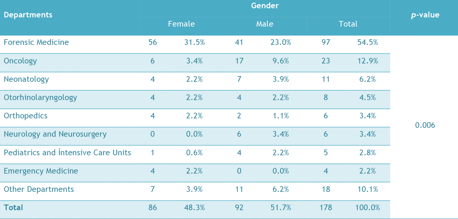
Aim: This study aimed to evaluate the conditions and indications in which a research and training hospital directs outpatient and inpatient consultations to an oral and maxillofacial surgery clinic and to evaluate the expectations of physicians in the treatment and management of various conditions. Methodology: The outpatient and inpatient consultations directed from Tokat Gaziosmanpaşa University’s clinics and internal and surgical medical sciences services to the same institution’s Oral and Maxillofacial Surgery Clinic between January 2013 and January 2022 were included. The patient characteristics, the aims of and responses to the consultations, and the applied interventions were recorded and classified. The quantitative data were analyzed with a t-test, and the classified data were analyzed with cross-tables and chi-square tests. P values below 0.05 were considered significant. Results: The medical records and consultation notes of 178 patients (92 males, 86 females) were evaluated. Of the consultations referred, 97 were requested from Forensic Medicine, 23 from Oncology, 11 from Neonatology, 8 from Otorhinolaryngology, 6 from Orthopedics, 6 from Neurology and Neurosurgery, 5 from Pediatrics, 4 from Emergency Medicine, and 18 from other departments. When the contents of the consultation were examined, it was observed that most of them were requested for age determination, post-assault examination, and dental abscesses. Conclusion: The present study indicates that an oral and maxillofacial surgery clinic is in close interaction with many medical branches and disciplines, especially forensic medicine, oncology, otorhinolaryngology, and neonatology. How to cite this article: Çolak S, Demirsoy MS, Erdil A, Keldal G, Altan A, Akbulut N. Evaluation of consultation contents directed from a university training and research hospital to an oral and maxillofacial surgery clinic. Int Dent Res 2023;13(1):35-43. https://doi.org/10.5577/idr.2023.vol13.no1.6 Linguistic Revision: The English in this manuscript has been checked by at least two professional editors, both native speakers of English.Consultation, dentistry, Forensic Odontology, Oncology, Oral and Maxillofacial SurgerySefa Çolak, Mustafa Sami Demirsoy, Aras Erdil, Göksal Keldal, Ahmet Altan, Nihat Akbulut30.04.202335-43 -
Ectodermal dysplasia: A retrospective evaluation of the clinical findings of forty-four cases in the 0-16 years age
Aim: The aim of this study was to review the craniofacial anomaly results of children diagnosed with ectodermal dysplasia (ED) and to identify the oral requirements of ED cases. Methodology: The data of this study were obtained by taking clinical examinations and radiographs on forty-four (44) children (22 females and 22 males), 0-16 years aged, who were admitted to the Dicle University, Faculty of Dentistry due to dental problems and were diagnosed with ED. The number of affected siblings was ascertained, and systemic findings were evaluated. Malformations in the hair, nails, nose, skin, lips, and teeth were clinically examined. Results: The most common clinical findings were sparse hair, dry skin, sweating problems, respiratory difficulty, saddle nose, a history of fever, hearing loss, and deformation in the nails. In the intraoral and radiological examinations, findings were evaluated of conical teeth, protuberant lips, prosthetic rehabilitation, impacted teeth, and abnormal root resorption. The rates of ED in the siblings of the ED cases were determined as 39.2% in the siblings of male cases and 37.9% in the siblings of female cases. Conclusion: The frequency rates of the anomalies seen in ED obtained in this study can be considered important as a guide for further studies of individuals with ED. When multiple missing teeth and conical teeth are encountered, the dental practitioner should investigate whether or not there are other symptoms of ED, and it must not be forgotten that the dentist may be the first step in the diagnosis of this genetic irregularity. How to cite this article: Akleyin E, Sarıyıldız Osmanoğulları C, Yavuz İ, Adıgüzel Ö. Ectodermal dysplasia: A retrospective evaluation of the clinical findings of forty-four cases in the 0-16 years age. Int Dent Res 2022;12(1):21-6. https://doi.org/10.5577/intdentres.2022.vol12.no1.4 Linguistic Revision: The English in this manuscript has been checked by at least two professional editors, both native speakers of English.ectodermal dysplasia, malformation, conical tooth, sparse hairEbru Akleyin, Cansu Osmanoğulları Sarıyıldız, İzzet Yavuz, Özkan Adıgüzel30.08.202221-26 -
Comparative evaluation of the color stability of universal composites
Aim: The aim of this study is to compare the color stability of different universal composites and to evaluate the effects of keeping them in water for 1 week before coloring. Methodology: Filtek Ultimate (FU), Estelite Ʃ Quick (EQ), and Ceram X One (CX) composites were used. The specimens were kept in the dark, dry environment for 24 hours, then polished, and each composite group was divided into 3 groups: the control, the group that was kept in water for 1 week before coloring and coloring (n=7). The samples in the control group were kept in distilled water for 3 weeks. Samples in the coloring group were colored with coffee for 2 weeks (C), and the samples in the last group were kept in water for 1 week and then stained with coffee for 2 weeks (W+C) (n=7). Color measurements for all samples were taken twice, once before and once after the coloring period. The color changes of the samples were measured using the ∆E* parameter, which was calculated with before and after measurements. Results: For all composite resins, the ΔE values of the samples in the W+C and C groups were significantly higher than those in the control group. When the composites were compared with each other, the differences between the ΔE values of the composites in the control, W+C, and C groups were not statistically significant. For FU and ESQ, at the end of the coloration period, there were no statistically significant differences between the W+C and C groups. However, for CX, the color change of the samples in the W+C group was significantly higher than that in the C group. Conclusion: Storage in distilled water for 1 week before staining did not reduce the color change values of the samples. Thus, after composite restorations are performed, there is no need to advise patients to avoid staining foods, especially in the first week after restoration. How to cite this article: Abdulateef OF, Çobanoğlu N, Gürses M. Comparative evaluation of the color stability of universal composites. Int Dent Res 2021;11(Suppl.1):234-7. https://doi.org/10.5577/intdentres.2021.vol11.suppl1.34 Linguistic Revision: The English in this manuscript has been checked by at least two professional editors, both native speakers of English.color stability, composite resin, spectrophotometry, ΔE value, ΔL valueMerve Gürses, Nevin Çobanoğlu, Omar Faez Abdulateef30.09.2021234-237 -
Evaluation of the push-out bond strength of ProRoot MTA and Bio-dentine after removal of calcium hydroxide and triple antibiotic paste
Aim: To evaluate the effect of calcium hydroxide (CH) and triple antibiotic paste (TAP) on the push-out bond strength of ProRoot® MTA and Biodentine®. Methodology: Root canals of 120 human teeth were instrumented using rotary files. To obtain a standard diameter of 1.5 mm, the roots were instrumented with a #6 Peeso reamer. The reamers passed 1 mm beyond the apex point to simulate open apices. The roots were randomly assigned into the following groups: Group 1 was administered calcium hydroxide (CH); Group 2 was administered triple antibiotic paste (TAP); and Group 3 was a control group and had no medicament. The medicaments were removed after 3 weeks. Thereafter, the specimens were divided into two subgroups (n = 20) according to which calcium silicate-based cement, ProRoot MTA (Dentsply Maillefer, Ballaigues, Switzerland) or Biodentine (Septodont, France), was applied. The push-out test was performed, and the data were analyzed statistically using one-way ANOVA and by Tukey’s post hoc test. Results: In both the ProRoot MTA and Biodentine groups, pairwise comparisons between medicaments showed similar values to the control group (p>0.05). Overall, there was a predominance of cohesive failures between root dentin and the cements. Conclusions: ProRoot MTA and Biodentine showed similar bond strength values, and prior application of TAP or CH did not affect the bond strength significantly. How to cite this article: Tulumbacı F, Erkmen Almaz M, Arıkan V, Mutluay MS. Evaluation of the push-out bond strength of ProRoot MTA and Biodentine after removal of Calcium Hydroxide and Triple Antibiotic Paste. Int Dent Res 2018;8(2):50-5. Linguistic Revision: The English in this manuscript has been checked by at least two professional editors, both native speakers of English.Biodentine, calcium hydroxide, mineral trioxide aggregate, ProRoot MTA, triple antibiotic pasteFatih Tulumbacı, Merve Erkmen Almaz, Volkan Arıkan, Merve Safa Mutluay27.08.201850-55 -
The determination of mercury levels in the blood and hair of rabbits with amalgam fillings
Purpose: This study was conducted to investigate the level of blood and hair mercury transition in rabbits depending on the number of amalgam filled teeth in the mouth. Material and Methods: A total of 18 male rabbits from New Zealand were used in the study. The rabbits were separated into three groups in this study. The first group was named as control group, the second group was named as dichotomous filled group, and the third group was named as quadruple amalgam filled group. Blood samples were collected from filler animals before the filling, at 24 hour, at the first week and month and also hair samples were taken before the filling and at the first month. The transfer of mercury, taken by the blood and feather samples of rabbits in the study, was made with the hydride system in the absorption spectrophometer. Results: The data’s, obtained from the study, were evaluated by using the SPSS 21 package program. Compared by the control group at the 24 th hour, any statically difference wasn’t seen between dichotomous and quadruple amalgam filling groups in terms of blood mercury level. (P>0.05). However, the level of blood mercury, one week and one month later was significantly higher than the control group.(P<0.05) In the all periods, the level of blood mercury was found similar between dichotomous and quadruple amalgam filling groups (P>0.05). Conclusions: Mercury couldn’t be detected in hair samples. As a result, it was revealed that amalgam filling affects the blood mercury level. How to cite this article: Yerebasan Ü, Servi K, Ataş O, Doğan MS. The determination of mercury levels in the blood and hair of rabbits with amalgam fillings. Int Dent Res 2020;10(1):12-16. https://doi.org/10.5577/intdentres.2020.vol10.no1.3 Linguistic Revision: The English in this manuscript has been checked by at least two professional editors, both native speakers of English.Mercury, amalgam, rabbit, blood, hairÜlkü Yerebasan, Kadir Servi, Osman Ataş, Mehmet Sinan Doğan30.04.202012-16 -
A rare case of an immature incisor with horizontal root fracture traumatized at the time of eruption
Dental trauma resulting in root fracture is a rare condition that affects up to 7% of permanent teeth, and injury to immature teeth is even rarer. This case report aimed to show the prognosis of a horizontal root-fractured immature maxillary incisor traumatized at the time of eruption. Because of an accident, a 6-year-old boy was referred to our clinic with horizontal root fracture of the permanent maxillary left central incisor associated with an extrusive luxation. The coronal segment was repositioned, and a dental splint was applied for 7 weeks. After 48 months, clinical examination revealed a positive response to electrical pulp testing and an absence color change of the tooth. Continuation of root development and calcification of the coronal pulp space was observed radiographically. It was concluded that fixation of teeth is a conservative treatment for immature teeth with horizontal root fractures, resulting in the pulp vitality and spontaneous healing with no other treatment. How to cite this article: Erkmen Almaz M, Şaroğlu Sönmez I, Akbay Oba A. A rare case of an immature incisor with horizontal root fracture traumatized at the time of eruption. Int Dent Res 2018;8(2):93-5. Linguistic Revision: The English in this manuscript has been checked by at least two professional editors, both native speakers of English.Child trauma, incisor, tooth eruption, horizontal root fractureMerve Erkmen Almaz, Işıl Şaroğlu Sönmez, Aylin Akbay Oba27.08.201893-95 -
Three-dimensional Finite Element Analysis of Endodontically Treated Tooth Restored with Carbon and Titanium Posts
Aim: This study compared the stress distributions of endodontically treated tooth restored with carbon and titanium post during different loading conditions. Methodology: A 3-dimensional finite element model was created to represent an endodontically treated maxillary central incisor tooth with its supporting structures. Stress distribution and stress values were then calculated by considering the three dimensional von Mises stress criteria. Results: A 100-N static vertical occlusal load was applied on the node at the center of occlusal surface of the tooth. The von Mises stress values for carbon post model was on the coronal third and the cervical area of the root in the range of 353.149 and 13.878 MPa, for titanium post model was 540.736 and 22.777 MPa. Conclusion: This study shows that the titanium post yields larger stresses than the carbon post. How to cite this article: Adıgüzel Ö, Kaya S, Yiğit Özer S, Değer Y, Göncü Başaran E, Yavuz İ. Three-dimensional Finite Element Analysis of Endodontically Treated Tooth Restored with Carbon and Titanium Posts. Int Dent Res 2011;2:55-59. Linguistic Revision: The English in this manuscript has been checked by at least two professional editors, both native speakers of English.Finite element analysis, carbon post, titanium postÖzkan ADIGÜZEL, Sadullah KAYA, Senem YİĞİT ÖZER, Yalçın DEĞER, Emine GÖNCÜ BAŞARAN, İzzet YAVUZ15.08.201155-59 -
The effect of final irrigation agents on push-out bond strength of calcium silicate-based cements to dentin
Aim: This aim of this study was to compare the effects of different chelating agents [ethylenediaminetetraacetic acid (EDTA), glycolic acid (GA) and citric acid (CA)] on the push-out bond strength (POBS) of two calcium silicate-based silicate cements (CSC) (Biodentine and PD MTA White). Methodology: Dentin discs of 1 ± 0.2 mm thickness were taken from the middle root region of thirty-nine extracted mandibular premolar teeth and two holes (1 mm diameter) were drilled in each disc (n = 78 holes). The samples were then randomly divided into three groups (n = 26 holes) according to the final irrigation agents: Group 1: 20% CA, Group 2: 17% EDTA, Group 3: 10% GA. Then, two different materials were applied to the holes in each group (n = 13 holes): a: PD MTA White, b: Biodentine. POBS test was performed at a crosshead speed of 0.5 mm/min. The dentin discs were examined under stereomicroscope (25×) to assess the bond failure type. Data were analyzed using a two-way analysis of variance (ANOVA) and comparison of main effects was examined with the Bonferroni test, and multiple comparisons were analysed with the Tukey HSD test. The level of signifcance was 5%. Results: Biodentine showed significantly higher POBS than PD MTA White ( p < 0.05). There was no significant difference between the final irrigation agents ( p > 0.05). CA – Biodentine group showed substantially higher POBS than EDTA – PD MTA White, CA – PD MTA White, and GA – Biodentine groups ( p < 0.05). Conclusion: While CA increased the POBS of Biodentine significantly, the POBS of PD MTA White was not affected by the final irrigation agents. How to cite this article: Yeniçeri Özata M, Falakaloğlu S, Batur M, Adıgüzel Ö, Goncharuk-Khomyn M. The effect of final irrigation agents on push-out bond strength of calcium silicate-based cements to dentin. Int Dent Res 2023;13(2):75-82. https://doi.org/10.5577/idr.2023.vol13.no2.5 Linguistic Revision: The English in this manuscript has been checked by at least two professional editors, both native speakers of English.Final irrigation agent, chelator, calcium silicate-based cement, push-out bond strength, Biodentine, PD MTA WhiteMerve Yeniçeri Özata, Seda Falakaloğlu, Muharrem Batur, Özkan Adıgüzel, Myroslav Goncharuk-Khomyn31.08.202375-82 -
The effect of immediate dentin sealing on fracture resistance and failure modes of resin composite onlay restorations
Aim: This study aimed to investigate the fracture resistance, failure modes, and reparability of molars restored with laboratory-processed resin composite (Ceramage, SHOFU Inc., Kyoto, Japan) onlays above or below the cemento-enamel junction (CEJ) in the presence or absence of immediate dentin sealing (IDS). Methodology: Forty extracted sound human molars were selected and divided into four groups: 1) Below CEJ with IDS, 2) Below CEJ without IDS, 3) Above CEJ with IDS, and 4) Above CEJ without IDS. Standardized mesio-occlusal (MO) onlay preparations were made with the reduction of mesio-buccal and mesio-palatinal cusps. Butt-joint preparation was used in the proximal box area extending 1 mm below the CEJ (Groups 1 and 2) or 1 mm above the CEJ (Groups 3 and 4). In Groups 1 and 3, IDS was applied immediately after tooth preparation. After cementation (Variolink N, Ivoclar Vivadent AG, Schaan, Liechtenstein), the specimens were thermally aged (20000 cycles, 5–55 ºC) and then subjected to load to failure (1 mm/min). The failure types and reparability of the onlays were recorded, and the data were analyzed using two-way ANOVA and Fisher’s exact tests. Results: The restorations in Group 4 showed the highest mean fracture strength compared to Group 1 ( p =0.038), Group 2 ( p =0.008), and Group 3 ( p =0.019). The interaction between the level of proximal extension and the presence of IDS did not influence fracture mode or reparability ( p >0.05). Conclusion: No difference in the fracture strength of resin composite onlays below CEJ with or without IDS was found; however, not using IDS for resin composite onlays above CEJ appears to be preferable. How to cite this article: Gözetici Çil B, Köymen SS, Ünal S, Dönmez N. The effect of immediate dentin sealing on fracture resistance and failure modes of resin composite onlay restorations. Int Dent Res 2024;14(1):27-33. https://doi.org/10.5577/intdentres.510Onlays, fracture strength, immediate dentin sealing, composite resinBurcu Gözetici Çil, Safiye Selin Köymen, Serpil Ünal, Nazmiye Dönmez30.04.202427-33 -
An evaluation of the relationship between dental anxiety and oral health in children based on maternal dental anxiety
Aim: This study aimed to understand the causes of dental anxiety that prevent optimal treatment in pediatric dental clinics and to examine the relationship between the child's dental anxiety level and the dental anxiety level of his or her parents. It also aimed to understand how the oral health of children whose parents have dental anxiety is affected by their parents’ feelings. Methodology: This study was conducted by evaluating the answers given to the Children’s Fear Survey Schedule-Dental Subscale (CFSS-DS) questionnaire, which was applied to 248 children without mental or developmental retardation who consulted the Dicle University, Faculty of Dentistry between January and October 2020. The Modified Dental Anxiety Scale (MDAS) questionnaire was applied to their parents. In addition, dmft+DMFT scores were determined by performing oral examinations on the children. Results: While a statistically significant relationship was found between the dental anxiety levels of the 248 children included in our study and the dental anxiety levels of their mothers, no significant relationship was found between the dental anxiety levels of their fathers and those of the children. Those children whose mothers had high dental anxiety scores had higher dmft+DMFT scores, compared to others involved in the survey. The dmft+DMFT scores of the children with high dental anxiety were found to be higher than those of other children. Conclusion: Parents, especially mothers, play an important role in the development of children’s dental anxiety. The fact that parents with dental anxiety often avoid taking their children to the dentist prevents necessary treatments from being performed on time and causes the children’s oral health to deteriorate over time. Informing parents about reducing dental anxiety and dentists’ efforts to teach coping strategies can be helpful for minimizing the impact of parents' dental concerns on their children. By increasing mothers’ awareness of oral and dental health, it can be ensured that both they and their children will benefit more from dentistry services. How to cite this article: Çelenk S, Kol Ş. An evaluation of the relationship between dental anxiety and oral health in children based on maternal dental anxiety. Int Dent Res 2021;11(Suppl.1):86-90. https://doi.org/10.5577/intdentres.2021.vol11.suppl1.14 Linguistic Revision: The English in this manuscript has been checked by at least two professional editors, both native speakers of English.dental anxiety, CFSS-DS, MDAS, maternal, questionnaireŞebnem Kol, Sema Çelenk30.09.202186-90 -
The effect of cold drinks on the coloration of resin-containing restorative materials
Aim: Resin-containing restorative materials are widely used in the restoration of teeth. Our aim in this study is to examine the color change of beverages consumed cold on resin-containing restorative materials. Methodology: Samples were prepared using composite resins and resin-based CAD/CAM blocks. All prepared samples were kept in cold tea, cold coffee, coke, energy drink, and distilled water after polishing. The color values of the samples were measured with a spectrophotometer at the beginning, on the 1st, 7th and 30th days, and the color change values were calculated with the CIEDE2000 formula. Statistical analysis of color change values was performed using two-way analysis of variance (ANOVA) and Tukey multiple comparison tests (p<0.05). Results: While there was no statistically significant difference in the color changes of the resin-containing dental materials at the end of the 1st day (p>0.05), at the end of the 7th and 30th days, cold tea caused the statistically highest color change (p<0.05). There was no statistically significant difference between the color change values of cold coffee, coke and energy drink on the materials in all time periods (p>0.05). Composite resins showed more color change than resin-based CAD/CAM blocks (p<0.05). Conclusion: Cold tea causes the most discoloration on resin-based materials. While the resin-containing CAD/CAM blocks showed a color change under the AT value in all cold drinks, the composite resins showed a color change above the AT value in cold tea and coffee. How to cite this article: Altıparmak ET, Aybala Oktay E, Karaoğlanoğlu S, Aydın N, Ersöz B, Özarslantürk S. The effect of cold drinks on the coloration of resin-containing restorative materials. Int Dent Res 2022;12(1):14-20. https://doi.org/10.5577/intdentres.2022.vol12.no2.3 Linguistic Revision: The English in this manuscript has been checked by at least two professional editors, both native speakers of English.CAD/CAM, composite resin, color change, cold drinksElif Tuğba Altıparmak, Elif Aybala Oktay, Serpil Karaoğlanoğlu, Numan Aydın, Bilge Ersöz, Savaş Özarslantürk30.08.202214-20 -
Comparative evaluation of the responses from ChatGPT-5, Gemini 2.5 Flash, and DeepSeek-V3.1 chatbots to patient inquiries about endodontic treatment in terms of accuracy, understandability, and readability
Aim: The aim of this study is to compare three large language models (LLMs) (ChatGPT-5, Gemini 2.5 Flash, and DeepSeek-V3.1) in terms of accuracy, understandability, and readability, based on the answers provided to frequently asked endodontic questions. Methodology: Thirty open-ended frequently asked questions were generated using the AlsoAsked and AnswerThePublic websites. Two experienced endodontists scored the accuracy of the responses using a 5-point Likert scale. The understandability of the responses was analyzed using the Patient Education Materials Assessment Tool for Printed Materials (PEMAT-P) tool. Readability was assessed using the Flesch-Kincaid Readability Score (FRES), Flesch-Kincaid Grade Level (FKGL), Gunning Fog Index (GFI), Simple Measure of Gobbledygook (SMOG), and Coleman-Liau Index (CLI). Group comparisons were performed using the ANOVA/Kruskal-Wallis test, followed by post-hoc Dunn-Bonferroni tests. Results: Inter-rater agreement was excellent (accuracy ICC: 0.908–0.917; reliability ICC: 0.992–0.995; all p < 0.001). A significant difference was found between the models in terms of accuracy (p < 0.001): DeepSeek-V3.1 (4.63 ± 0.81) scored the highest and performed significantly better than ChatGPT-5 (3.93 ± 0.79) and Gemini 2.5 Flash (3.67 ± 0.76). No significant difference between ChatGPT-5 and Gemini 2.5 Flash (p > 0.05). The understandability (PEMAT-P) scores were similar (p = 0.683), and all models scored above 70% (ChatGPT-5, 77.46%; Gemini, 76.04%; DeepSeek-V3.1, 77.57%). Differences were found in readability metrics: DeepSeek-V3.1 scored higher than ChatGPT-5 in FRES (p = 0.044); Gemini scored higher than DeepSeek-V3.1 in FKGL (p = 0.001); in GFI, Gemini 2.5 Flash scored higher than both ChatGPT-5 (p = 0.036) and DeepSeek-V3.1 (p < 0.001); in SMOG, Gemini outperformed DeepSeek-V3.1 (p = 0.003); and in CLI, ChatGPT-5 was higher than DeepSeek-V3.1 (p = 0.004). No significant correlation was found between readability and understandability (p > 0.05). Conclusion: DeepSeek-V3.1 outperformed ChatGPT-5 and Gemini 2.5 Flash in terms of accuracy. While all models produced similar scores above the PEMAT-P understandability threshold (70%), there were significant differences in readability metrics; furthermore, no model consistently reached the recommended 6 th –8 th grade level.Artificial intelligence, large language models (LLMs), ChatGPT, Gemini, DeepSeek, patient questions in endodontics, accuracy, understandability, PEMAT-P, readabilityMakbule Taşyürek, Özkan Adıgüzel, Mustafa Gündoğar, Myroslav Goncharuk-Khomyn, Hatice Ortaç31.12.2025123-135 -
The relationship between xerostomia and periodontal status in patients with cardiovascular diseases and type 2 diabetes
Aim: The aim of this cross-sectional study was to examine the correlation between patients' subjective perception of xerostomia and their periodontal status and to examine the potential association of this correlation with cardiometabolic disorders. Methodology: A total of 147 patients (40 systemically healthy, 33 with cardiovascular disease [CVD], 40 with type 2 diabetes, and 34 with cardiometabolic disease [CVD + diabetes]) who came to Necmettin Erbakan University, Faculty of Dentistry, Department of Periodontology for periodontal treatment and agreed to participate were included in the study. Sociodemographic information, tooth brushing frequency, systemic disease status, and clinical periodontal parameters (plaque index [PI], gingival index [GI], bleeding on probing [BOP], and mean pocket depths) were recorded. Patients were asked to complete an 11-item subjective xerostomia inventory questionnaire. Results: In the findings of our study, there was no statistically significant correlation between xerostomia total score and mean pocket depths, PI, GI, and BOP ( p >0.05). There was no statistically significant difference between the mean values of xerostomia total score according to systemic disease groups ( p =0.654). There is a statistically significant difference between the mean values of xerostomia total score according to periodontal status groups ( p =0.002). The mean xerostomia total score was 20.19 in periodontally healthy patients, 24.42 in gingivitis patients, and 22.73 in periodontitis patients. Conclusion: Although the results suggest that subjective dry mouth findings are associated with gingivitis, they do not affect pocket depth and do not increase susceptibility to periodontitis. Clinicians should emphasize the risk of gingivitis in patients with dry mouth and the importance of prevention. How to cite this article: Babayiğit O, Ögütcen Ö, Özkan Şen D, Taştan Eroğlu Z, Uçan Yarkaç F. The relationship between xerostomia and periodontal status in patients with cardiovascular diseases and type 2 diabetes. Int Dent Res. 2024; 14(S1):21-27. https://doi.org/10.5577/indentres.556Cardiometabolic diseases, gingivitis, periodontal status, xerostomia, type 2 diabetesOsman Babayiğit, Ömer Ögütcen, Dilek Özkan Şen, Zeynep Taştan Eroğlu, Fatma Uçan Yarkaç31.12.202421-27 -
The effect of different herbal teas on the color stability of nanohybrid and bulk-fill composites
Aim: The aim of this study is to investigate the effects of green tea and rosehip tea, on the color stability of nanohybrid and bulk-fill composite resins. Methodology: A total of 60 disc-shaped samples were made from two types of A2 resin with nanohybrid (Tetric N Ceram, Ivoclar Vivadent, Schaan, Liechtenstein) and bulk-fill (Filtek Bulk-Fill, 3M ESPE, MN, USA) structure with the help of 10 mm diameter and 2 mm thick Teflon molds in this study. All samples were finished and polished with 4-stage (thick, medium, fine, superfine) polishing discs (Sof-lex, 3M ESPE, MN, USA) and kept in an oven at 37 °C for 24 hours. The samples, whose initial color measurements were made by spectrophotometer, were divided into subgroups for the control group and two different herbal teas (Green tea, Rosehip) (n=10). Color measurements were repeated, and ΔE values were calculated. Wilcoxon, Kruskal-Wallis, and Bonferroni tests were used to analyze the data (p < 0.05). Results: There was no statistically significant difference in color change between the composite resins ( p > 0.05). No significant color change was found for either the Tetric N-Ceram or Filtek Bulk-Fill composite resin samples immersed in distilled water ( p > 0.05, ΔE<3.3). Conclusion: Rosehip and green tea affect the color stability of composite resin restorations. Whereas the color change caused by green tea is at a clinically acceptable level, that stemming from rosehip tea falls within clinically unacceptable levels. How to cite this article: Tunçdemir MT, Dereli Z, Bahar A. The effect of different herbal teas on the color stability of nanohybrid and bulk-fill composites. Int Dent Res 2022;12(Suppl.1):103-7. https://doi.org/10.5577/intdentres.445 Linguistic Revision: The English in this manuscript has been checked by at least two professional editors, both native speakers of English.Herbal tea, green tea, rosehip tea, nanohybrid, bulk-fill composite resin, color stabilityMakbule Tuğba Tunçdemir, Zeynep Dereli, Ayşe Bahar31.12.2022103-107 -
Treatment of Large Periapical Lesions without Surgical Approach: Report of Three Cases
The aim of this paper is to present the non-surgical management of three cases with large periapical lesions. Surgical treatment of large periapical lesions is often subject to various complications. Therefore, a non-surgical approach should be considered for the management of these lesions. The first was a maxillary lateral incisor with a periapical lesion of endodontic origin in a 19-year-old female patient, the second affected a maxillary central incisor in a 21-year-old female patient and the third was a mandibular molar in a 36-year-old male patient. The canals were prepared using crown down technique. During root canal treatment, sodium hypochlorite was used for irrigation and calcium hydroxide was used for the intra-canal dressing. Clinical evaluation was performed after 10 days. The teeth were asymptomatic and the root canals were obturated using lateral compaction technique. The patients were recalled at 3, 6 and 12 months. These radiolucent periapical lesions were treated successfully without surgical approach. How to cite this article: Arslan H, Karataş E, Barutcugil Ç, Topçuoğlu HS, Aladağ H. Treatment of Large Periapical Lesions without Surgical Approach: Report of Three Cases. Int Dent Res 2012;2:17-22 Linguistic Revision: The English in this manuscript has been checked by at least two professional editors, both native speakers of English.Non-surgical treatment, periapical lesion, root canal treatmentHakan Arslan, Ertuğrul Karataş, Çağatay Barutcugil, H. Sinan Topçuoğlu, Halit Aladağ15.04.201217-22 -
Evaluation of mental foramen localization in children by age and gender using cone-beam computed tomography
Aim: This retrospective study was carried out to obtain detailed data on mental foramen localization differences, which can guide dental practices such as endodontic treatments, surgical procedures, and dental anesthesia, preventing complications. Cone-beam computed tomography (CBCT) images of individuals aged 7–18 were analyzed according to gender and age. Methodology: The CBCT images of 120 individuals who met the research criteria were divided into six groups according to age, and 240 hemimandibles were measured. The gender distribution of the patients in each group was equal. Measurements were obtained using 3D volumetric images obtained via CBCT and the RadiAnt DICOM Viewer. When examining the differences between the groups, the Mann–Whitney U test was used for paired groups, and the Kruskal–Wallis H test and post hoc multiple comparison tests were used for groups of more than two. Results: Statistically significant differences in the parameters related to mental foramen localization were determined between the gender and age groups ( p <0.05). No statistically significant differences were found between the right and left sides of the individuals in all measurements ( p >0.05). Further, the mean values of the anatomical points were determined, which can be used to guide pediatric and general dentistry practices. Conclusion: In the present study, the mean values of the anatomical points measured were determined. Our study suggests that these values can be used as a guide in pediatric and general dentistry applications, such as local anesthesia, endodontic treatments, and surgical operations.mental foramen, anatomical landmark, cone-beam computed tomography, RadiAnt DICOM viewerHatice Kübra Başkan, Emin Caner Tümen30.04.202510-17 -
The Effects of Antioxidant Application and Time Factor on Fiber Post Bonding to Root Dentin after Intracoronal Bleaching
Aim: The aim was to evaluate the effects of antioxidant application and delayed cementation on the bond strength of fiber posts after intracoronal bleaching. Methodology: Fifty-five maxillary central incisors were used. Root canals were enlarged using Reciproc system up to R40 instrument. Root canals were irrigated, dried with paper-points, and obturated with gutta-percha and a sealer. In 44 teeth, root canal fillings were removed 2-mm coronally and canal orificies were sealed with glass-ionomer-cement. A 37% CP gel was used to bleach the teeth. Five experimental groups were defined (n=11). G1: No bleaching, G2: Immediate FPC, G3: Immediate FPC after SA application, G4: 14-days delayed FPC, G5: 14-days delayed FPC after SA application. Push-out tests were performed. Statistical interpretations were made (α=0.05). Results: There was no significant difference among the groups in apical thirds (p>0.05). However, significant differences were detected among the groups in middle and coronal thirds (p<0.05). Conclusion: The 14-days delayed cementation seems to be a more reliable method than the SA application before FPC procedure after intracoronal bleaching. How to cite this article: Aslan T, Üstün Y, Sağsen B, Şener İ, Biricik E, Tatlı Ş. The effects of antioxidant application and time factor on fiber post bonding to root dentin after intracoronal bleaching. Int Dent Res 2018;8(1):22-7. Linguistic Revision: The English in this manuscript has been checked by at least two professional editors, both native speakers of English.Internal bleaching, devital bleaching, antioxidant application, sodium ascorbate, delayed cementation, push-outTuğrul Aslan, Yakup Üstün, Burak Sağsen, İbrahim Şener, Eda Biricik, Şifa Tatlı30.04.201822-27


Open Access articles are distributed under the terms of the