Skip to main content Skip to main navigation menu Skip to site footer
logo
  • Login
  • Register
E-ISSN: 2146-1767
  • Home
  • Current Issue
  • Archives
  • For Authors
    • Author Guidelines
    • Online Submission
    • Reference Style
    • Call for Papers
    • Language Editing Service
  • For Reviewers
    • Reviewer Guidelines
    • Become a Reviewer
    • Reviewer Resources
  • Ethics & Policies
    • Artificial Intelligence (AI) Policy
    • Authorship and Contributorship
    • Conflicts of Interest
    • Copyright and Permission
    • Corrections and Retractions
    • Crossmark Policy
    • Editorial Policy
    • Human and Animal Rights
    • Informed Consent Policy
    • Journal Management
    • Open Access Statement
    • Research Ethics Policy
    • Publication Ethics Policy
  • Journal Info
    • About
    • Abstracting & Indexing
    • Peer Review Process
    • Publication Fee
    • Society Information
    • Useful Links
    • Contact
  1. Home /
  2. Search

Search

Advanced filters
Published After
Published Before

Search Results

##search.searchResults.foundPlural##
  • A Fibre-Reinforced Fixed Partial Denture on a Hemisectioned Tooth: A Case Report

    In modern dentistry, fibre-reinforced fixed dental prostheses are considered a useful alternative to classical metal-ceramic restorations. This method allows a conservative approach for replacing missing teeth that overcomes some of the drawbacks of conventional prostheses. Our patient required extraction of tooth #46 because of an apical lesion of the mesial root, and underwent extraction by hemisection. After healing, using the superior properties of the combined fibre/composite, an adhesive bridge restoration was applied with support from the distal root of tooth #46 and teeth #45 and #47.  How to cite this article:  Agüloğlu S, Ayna E, Özdemir E. A Fibre-Reinforced fixed partial denture on a hemisectioned tooth: A Case Report.  Int Dent Res 2011;1:38-41. Linguistic Revision:  The English in this manuscript has been checked by at least two professional editors, both native speakers of English.
    Fibre-reinforced, adhesive bridge, hemisection
    Süleyman AGÜLOĞLU, Emrah AYNA, Eylem ÖZDEMİR
    15.04.2011
    38-41
    Abstract Views: 1212 Download: 677 DOI 10.5577/intdentres.2011.vol1.no1.7
  • Comparison of the effect of different rapid maxillary expansion treatments on tooth hardness values: Splint mouth study

    Aim: The purpose of this study, planned as a splint mouth design, is to compare the effects of different rapid maxillary expansion (RME) appliances on the hardness values of teeth. Methodology: This study utilized extracted upper first premolar teeth from a previous study conducted by Yıldırım and Akın in 2019 on 20 patients. Five patients were excluded from this study due to fractures of the tooth crown during extraction, which also necessitated the exclusion of the opposing arch teeth. The extracted teeth were stored in distilled water at +4 degrees Celsius. The crowns and roots of these teeth were separated at the cementoenamel junction using an aerator and a diamond bur under water cooling. The crowns were then embedded into rings of standard thickness using the cold acrylic dripping method. Hardness measurements were performed using a Vickers Hardness tester. Enamel samples obtained from the patients were placed under a test microscope and examined at X40 magnification; force was applied to each enamel sample for 10 s. The device automatically calculated and recorded the microhardness value relationship. The Shapiro-Wilk test was used to confirm the normal distribution of the data. Comparisons between groups were conducted using Student's t-test. Results: The group using the hybrid maxillary expansion appliance showed lower hardness values than the conventional group, although the difference was not statistically significant. Conclusion: Fixed orthodontic treatments similarly increase the demineralization of teeth. Therefore, individual or preventive measures by dentists or orthodontists should be taken against mineral losses.   How to cite this article: Sağoğlu S, Yıldırım M, Akın M, Erdur EA. Comparison of the effect of different rapid maxillary expansion treatments on tooth hardness values: Splint mouth study. Int Dent Res. 2024;14(S1):45-49. https://doi.org/10.5577/indentres.564
    Hardness test, rapid maxillary expansion, splint mouth design, tooth extraction, hardness
    Seda Sağoğlu, Mücahid Yıldırım, Mehmet Akın, Emire Aybüke Erdur
    31.12.2024
    45-49
    Abstract Views: 2114 Download: 474 DOI 10.5577/indentres.564
  • Evaluation of changes in alveolar bone remodeling after orthodontic treatment using fractal analysis: A retrospective study

    Aim: The aim of this retrospective study was to analyze trabecular changes in alveolar bone remodeling after orthodontic treatment by evaluating dental panoramic radiographs using the fractal dimension (FD) analysis method. Methodology: This study included 146 individuals with dental class II malocclusion who had undergone fixed orthodontic treatment. The mean age of the patients was 14.88±2.11 years. Patients were divided into three groups: nonextraction control group (Group 1, n=45), moderate anchorage group with extraction (Group 2, n=53), and maximum anchorage group with extraction (Group 3, n=48). The region of interest in the maxillary first premolar apical area was determined using pre- and post-treatment (T0 and T1) panoramic radiographs obtained from archival records. FD analysis was conducted using ImageJ software (version 1.53, NIH). For intragroup data comparison, the paired sample t-test and Wilcoxon test were used. For intergroup data comparison, the Kruskal–Wallis and Mann–Whitney U tests were used. Statistical significance was set at p <0.05. Results: No significant difference in FD values was found between the groups at T0 ( p >0.05). At T1, compared to T0, FD values showed a significant decrease in Groups 2 and 3 (p<0.05) and no significant change in Group 1 ( p >0.05). At T1, FD values of Groups 2 and 3 were significantly lower than those for Group 1 (p<0.05); no significant difference was found within Groups 2 and 3 (p>0.05). T0/T1 FD change was significantly more in Groups 2 and 3 compared to Group 1 ( p <0.05). Conclusion: Although fixed orthodontic treatments with extraction caused significant changes in the fractal dimensions of alveolar trabecular structures when compared to nonextraction, they did not cause destructive changes.   How to cite this article: Çiçek O, Yılmaz H, Melayim H. Evaluation of changes in alveolar bone remodeling after orthodontic treatment using fractal analysis: A retrospective study. Int Dent Res 2024;14(1):19-26. https://doi.org/10.5577/intdentres.517  
    Fractal analysis, bone remodeling, fixed orthodontic treatment, tooth extraction, trabecular structure
    Orhan Çiçek, Hakan Yılmaz, Hamza Melayim
    30.04.2024
    19-26
    Abstract Views: 2471 Download: 763 DOI 10.5577/intdentres.517
  • Effect of three different maxillary expansion appliances on root resorption

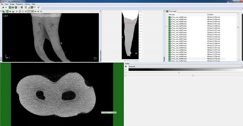
    Aim: The study was to examine rapid maxillary expansion (RME), semi-rapid maxillary expansion (SRME), and slow maxillary expansion (SME) compare the root resorptions occurring in the supporting upper first premolar teeth during treatment by using micro-computed tomography (micro-CT). Methodology: Thirty individuals that required maxillary expansion and upper first premolar tooth extraction were divided into three groups consisting of 10 individuals for RME, SRME, and SME. RME group, first premolar and first molar teeth with hyrax screw were activated ¼ tour twice daily with the banded hyrax appliances. SRME group, acrylic-bonded hyrax appliances were activated ¼ tour per morning and night until the opening of the suture. Upon the opening of the suture, it was activated ¼ tour per every two days until sufficient maxillary expansion was gained. SEM group, quad helix appliances were activated every 20 days. After 12 weeks of the retention period, the upper first premolar teeth were extracted. The teeth were scanned with Micro-CT (SkyScan 1172, Kontich, Belgium), and images were analyzed with a Dell Precision T5500 to determine the resorption crater volumes. Results: In all samples, the resorption craters were concentrated on the buccal surfaces of the tooth roots. The highest resorption craters volume, the total resorption volume, was observed in the RME group, followed by the SRME group, and the least resorption crater volume was detected in the SME group. Conclusion: Since the supporting teeth were exposed to more force on the root surfaces, the most resorption was seen in the RME group.   How to cite this article: Topal R, Gündüz Arslan S. Effect of three different maxillary expansion appliances on root resorption. Int Dent Res 2023; 13(1):19-26. https://doi.org/10.5577/idr.2023.vol13.no1.4   Linguistic Revision:  The English in this manuscript has been checked by at least two professional editors, both native speakers of English.
    Root resorption, maxillary expansion, expansion speed, palatal expansion, micro-computed tomography
    Refika Topal, Seher Gündüz Arslan
    30.04.2023
    19-26
    Abstract Views: 1857 Download: 534 DOI 10.5577/idr.2023.vol13.no1.4
  • Ozone applications in dentistry: A review

    Ozone (O 3 ) therapy has been applied in medicine and dentistry for many years and has become increasingly widespread. Ozone, composed of three oxygen atoms (O 3 ), is a strong antioxidant and a highly effective antimicrobial agent. Ozone gas has high oxidizing properties and exhibits a 1.5-fold higher antimicrobial effect than chloride. Its use has become popular in many fields of dentistry in recent years, owing to its various properties. It is categorized as one of the non-invasive, atraumatic, protective, and preventive treatment modalities that have been improved by modern dentistry, and its acceptability among patients has increased as a result. Ozone is an antimicrobial, analgesic, and biosynthetic agent that stimulates the immune system, enhances blood circulation capacity, and positively affects oxygen transport mechanisms. It influences both cellular and humoral immunity. All three forms of ozone (gaseous, oil, and solution) can be used in dentistry. It is indicated for the treatment of approximately 300 different pathological conditions. Ozone is used in endodontics (irrigation solution, intracanal medicament, abscess cases, disinfection), periodontology (gingivitis, periodontitis, periimplantitis, surgical injuries, prophylaxis), oral pathology (stomatitis, aphthous ulceration, candidiasis, herpes lesions), oral, dental, and jaw surgery (implantation, reimplantation, simple-to-complicated tooth extraction, homeostasis, wound healing, temporomandibular joint (TMJ) surgery, osteonecrosis cases), prosthetic dental treatment (disinfection of fixed and removable prostheses), restorative dental treatment (caries lesions, dentine hypersensitivity, cracked tooth, bleaching), orthodontics (remineralization and prophylaxis of white spot lesions), pedodontics (caries lesions, root canal treatment, positive psychological impact, short treatment duration), and sterilization of dental instruments and units. This review aimed to describe the usage fields and effects of ozone in dental applications, provide insights into clinical practices in other fields of dentistry, and introduce newly conducted in vitro and in vivo studies.
    Ozone, ozone therapy, dentistry, disinfection
    Merve Doğan İlik, Özkan Adıgüzel, Myroslav Goncharuk-Khomyn
    31.12.2025
    222-236
    Abstract Views: 1750 Download: 609 DOI 10.5577/intdentres.730
  • Impact of COVID-19 pandemic on pediatric dentistry treatment procedures

    Aim: The SARS-CoV-2 (COVID-19) pandemic has forced pediatric dentistry to limit clinical procedures due to children’s role in spread of the virus and transmission routes in pediatric patients. In order to minimize contamination and cross-infection risk, dental examinations and treatments had been postponed and patient admission protocols rearranged. This study aimed to evaluate the effects of pandemic on the number of patients admitted and treated in the pediatric dentistry clinic of a university hospital in the one-year period following the March 2020 global pandemic declaration. Methodology: The study included pediatric patients aged 0-14 years admitted to a pediatric dentistry clinic of a faculty of dentistry between March 2019 and March 2021 in order to compare and make a retrospective evaluation of the one-year time intervals before and after the pandemic announcement in March 2020. Children were divided into the age groups 2-6 and 7-14. Evaluation parameters were chosen as examination, radiograph (panoramic and periapical), fissure sealing or topical fluoride application, compomer, composite filling, pulpotomy, root canal treatment, prefabricated crown, tooth extraction, and space maintainer. Results: The results of the study showed a statistically significant decrease in monthly average number of examinations after the declaration of the pandemic regardless of age and gender (p<0.001). In both age groups, there is a proportional decrease in pulpotomy, composite filling, fissure sealant or topical fluoride application, root canal treatment, compomer filling, prefabricated crown procedures during the pandemic compared to the pre-pandemic, while the number of filming procedures increased proportionally (p<0.001). Conclusion: Dental clinicians were advised to take on more radical treatment options during the pandemic. Further studies are needed to examine the effects of the statistical decrease in the number of procedures in the field of pediatric dentistry and treatment services on long-term community oral health.   How to cite this article: Yüksel BN, Bezgin T. Impact of COVID-19 pandemic on pediatric dentistry treatment procedures. Int Dent Res 2021;11(3):140-8. https://doi.org/10.5577/intdentres.2021.vol11.no3.2   Linguistic Revision:  The English in this manuscript has been checked by at least two professional editors, both native speakers of English.  
    COVID-19 pandemic, dental treatment, pediatric dentistry, retrospective study
    Burcu Nihan Yüksel, Tuğba Bezgin
    31.12.2021
    140-148
    Abstract Views: 2923 Download: 690 DOI 10.5577/intdentres.2021.vol11.no3.2
  • Evaluation of staining susceptibility of teeth after office bleaching

    Aim: The aim of this study was to evaluate the discoloration effect of different beverages on previously office-bleached teeth. Methodology: Forty upper incisor teeth with indication for extraction due to periodontal reasons were used in this study. Teeth with discoloration, fractures, caries, or restorations on the crown surface were excluded. Before applying the bleaching agent to the enamel surface, the teeth were embedded into acrylic resin. Two office bleaching sessions were applied. Teeth were randomly divided into five experimental groups (n=8): tea, coffee, cola, and milk, with artificial saliva as the control. The teeth were soaked in beverages for 5 minutes a day for 1 month and were kept in artificial saliva for the rest of the time. Color measurements were taken using a spectrophotometer and the L, C, h values ​​were recorded. Initial color measurements were taken after bleaching sessions. Color measurements were repeated for each beverage after soaking for 24 hours, 72 hours, 1 week, and 1 month and color changes (ΔE 00 ) were calculated using the CIEDE2000 formula. Data were analyzed by two-way repeated analysis of variance using the IBM SPSS 25.0 program ( p <0.05). Results: Teeth kept in coffee and tea revealed significantly different mean ΔE 00 values ​​in the comparisons between the 1st month and 24th hour and between the 72nd hour and 1st week ( p <0.001, p <0.001, and p =0.002, respectively). During inter- and intragroup comparisons of the milk and cola groups, no significant differences were detected at examination intervals ( p >0.05). In the 1st month measurements, the mean ΔE 00 values ​​obtained from samples soaked in coffee and tea were statistically higher than the artificial saliva group ( p =0.006 and p =0.007). The mean ΔE 00 values ​​obtained at the 24th and 72nd hour and at the 1st week did not differ significantly among the tested beverages ( p >0.05). Conclusion: After bleaching, discoloration increased over time. Tea and coffee caused the most significant discoloration.   How to cite this article: Çam M, Kütük Ömeroğlu M. Evaluation of staining susceptibility of teeth after office bleaching. Int Dent Res. 2024;14(S1):65-69. https://doi.org/10.5577/indentres.550
    Discoloration, staining, hydrogen peroxide, tooth whitening, office bleaching, CIEDE2000
    Melek Çam, Merve Kütük Ömeroğlu
    31.12.2024
    65-69
    Abstract Views: 1629 Download: 464 DOI 10.5577/indentres.550
  • Mandibular Buccal Bone Thickness In Southeastern Anatolian People: A Cone-Beam Computed Tomography Study

    Aim: Buccal bone thickness is an important factor in implant treatment, bone health after tooth extraction, apical surgery, and esthetic outcomes. We evaluated the distance from the mandibular premolar and molar teeth apices to the buccal cortical bone in southeastern Anatolian people using cone-beam computed tomography (CBCT). Methodology: This retrospective study was performed in 461 posterior teeth (220 premolars, 241 molars) of 133 patients (62 females, 64 males) at Dicle University, Faculty of Dentistry, Department of Oral and Maxillofacial Radiology. Data were analyzed using Student’s t-tests and Tukey HSD tests. Results: The mandibular buccal bone was thicker in men than in women, but the difference was not statistically significant (p>0.05). The thinnest point of the mandibular buccal bone was measured in women as 2.431 mm and in men as 2.491 mm in the first premolar teeth. The thickest point of the mandibular bone was measured in women as 7.940 mm and in men as 7.859 mm in the distal roots of mandibular second molar teeth. For the mandibular first and second premolars, there was no significant difference in buccal bone thickness among the 10–29-, 30–49-, and 50–69-year age groups. The difference between the first and second molar mesial and distal roots of the age groups was significant at the level of buccal root thickness (p<0.05). Conclusions: It is important to examine buccal bone thickness (with CBCT) before surgical dental procedures for appropriate implant planning and surgical endodontic treatment.  How to cite this article:  Aktuna Belgin C, Adiguzel O, Bud M, Colak M, Akkus Z. Mandibular Buccal Bone Thickness In Southeastern Anatolian People: A Cone-Beam Computed Tomography Study. Int Dent Res 2017;7:6-12. Linguistic Revision:  The English in this manuscript has been checked by at least two professional editors, both native speakers of English.
    Buccal bone thickness, cone-beam computed tomography, mandible, alveolar bone
    Ceren Aktuna Belgin, Ozkan Adiguzel, Marius Bud, Mehmet Colak, Zeki Akkus
    15.04.2017
    6-12
    Abstract Views: 3159 Download: 1121 DOI 10.5577/intdentres.2017.vol7.no1.2
1 - 8 of 8 items

Journal Issue

Vol. 16 (2026): International Dental Research
Vol. 16 (2026) Continuous Publication
View Current Issue

Published Volumes

2026
  • Vol. 16 (2026): International Dental Research
  • Vol. 16 No. S1 (2026): International Dental Research
  • Vol. 16 No. Advanced Online (2026): International Dental Research
2025
  • Vol. 15 No. 3 (2025): International Dental Research
  • Vol. 15 No. 2 (2025): International Dental Research
  • Vol. 15 No. 1 (2025): International Dental Research
2024
  • Vol. 14 No. 3 (2024): International Dental Research
  • Vol. 14 No. Suppl. 1 (2024): International Dental Research
  • Vol. 14 No. 2 (2024): International Dental Research
  • Vol. 14 No. 1 (2024): International Dental Research
  • 2024: Advanced Online
2023
  • Vol. 13 No. S1 (2023): International Dental Research
  • Vol. 13 No. 3 (2023): International Dental Research
  • Vol. 13 No. 2 (2023): International Dental Research
  • Vol. 13 No. 1 (2023): International Dental Research
2022
  • Vol. 12 No. Suppl. 1 (2022): International Dental Research
  • Vol. 12 No. 3 (2022): International Dental Research
  • Vol. 12 No. 2 (2022): International Dental Research
  • Vol. 12 No. 1 (2022): International Dental Research
  • No. Accepted Articles: International Dental Research
Show more Show less
2021
  • Vol. 11 No. 3 (2021): International Dental Research
  • Vol. 11 No. Suppl. 1 (2021): International Dental Research
  • Vol. 11 No. 2 (2021): International Dental Research
  • Vol. 11 No. 1 (2021): International Dental Research
2020
  • Vol. 10 No. 3 (2020): International Dental Research
  • Vol. 10 No. 2 (2020): International Dental Research
  • Vol. 10 No. 1 (2020): International Dental Research
2019
  • Vol. 9 No. 3 (2019): International Dental Research
  • Vol. 9 No. 2 (2019): International Dental Research
  • Vol. 9 No. 1 (2019): International Dental Research
2018
  • Vol. 8 No. 3 (2018): International Dental Research
  • Vol. 8 No. 2 (2018): International Dental Research
  • Vol. 8 No. 1 (2018): International Dental Research
2017
  • Vol. 7 No. 3 (2017): International Dental Research
  • Vol. 7 No. 2 (2017): International Dental Research
  • Vol. 7 No. 1 (2017): International Dental Research
2016
  • Vol. 6 No. 1 (2016): Volume 6, No 1, 2016
2012
  • Vol. 2 No. 3 (2012): Volume 2, No 3, 2012
  • Vol. 2 No. 1 (2012): Volume 2, No 1, 2012
  • Vol. 2 No. 2 (2012): Volume 2, No 2, 2012
2011
  • Vol. 1 No. 3 (2011): Volume 1, No 3, 2011
  • Vol. 1 No. 2 (2011): Volume 1, No 2, 2011
  • Vol. 1 No. 1 (2011): Volume 1, No 1, 2011

socialshare

Submit Your Paper
Become a Reviewer

sidebarmenu

Journal Menu

 Home

 Aims & scopes

 Editorial Board

 Author Guidelines

 Abstracting & Indexing

 Peer Review Process

 Ethics Statement

 Plagiarism Policy

 Digital Archiving Policy

 Corrections and Retractions

 Journal History

 Journal Statistics

 Society Information

 Congresses & Events

 Language Editing

 Contact

idr


Citations
Updated daily Google Scholar
All Since 2021
Citations 1073 873
h-index 13 11
i10-index 18 12
0 50 100 150 200 250
  • 37 2019
  • 50 2020
  • 71 2021
  • 112 2022
  • 146 2023
  • 140 2024
  • 202 2025
  • 199 2026
Citations per year
Year Citations
2019 37
2020 50
2021 71
2022 112
2023 146
2024 140
2025 202
2026 (to date) 199

2026 counts citations recorded so far this year.

icmje and ithenticate

Indexed in CiteLink

EBSCO

TR Dizin Journal List

Keywords

Information

  • For Readers
  • For Authors
  • For Librarians

Latest publications

  • Atom logo
  • RSS2 logo
  • RSS1 logo
Most Read Articles
  • Endodontic Irrigation Solutions: A Review
    13336 13336 views 2017
  • Mechanical properties of resin composites containing bioactive glass and experimental nano zinc-silica complex
    11127 11127 views 2021
  • Areas for use of PEEK material in dentistry
    9560 9560 views 2018
  • Comparative evaluation of the responses from ChatGPT-5, Gemini 2.5 Flash, and DeepSeek-V3.1 chatbots to patient inquiries about endodontic treatment in terms of accuracy, understandability, and readability
    7864 7864 views 2025
  • Evaluation of the color stability and surface roughness of dual-cure, bulk-fill composites
    7181 7181 views 2021

announcement

Announcement

visitors

Visitors 

Flag Counter

Web Analytics

View My Stats

Most Cited Articles
  • Areas for use of PEEK material in dentistry
    162 162 citations 2018
  • Contemporary Permanent Luting Agents Used in Dentistry: A Literature Review
    61 61 citations 2011
  • The Latest Developments Related to Composite Resins
    44 44 citations 2017
  • PEEK in dentistry, properties and application areas
    37 37 citations 2020
  • A Literature Review of Self Adjusting File
    31 31 citations 2011

Browse

  • Categories
    • Open Access
    • Original Article
    • Review
    • Case Report
    • Letter to Editor
logo
E-ISSN: 2146-1767

Download App

Contact

Publisher: International Dental Research

Email: info@dental-research.com

Website: www.dental-research.com

Address: Publisher ZEUS PUBLISHING www.zeuspublishing.com

Social Media

ElsevierCiteLinkEBSCOOpen AccessCrossrefGoogle ScholarCOPECreative Commons

Copyright © 2011-2026 | International Dental Research. All rights reserved.
CC BY Open Access articles are distributed under the terms of the Creative Commons Attribution 4.0 International License,
which permits unrestricted use, distribution, and reproduction provided the original work is properly cited.

Privacy notice

This journal stores a session cookie that is required for the site to work, and keeps usage statistics that do not identify you.

More information