Examination of root canal morphology of teeth affected by Molar Incisor Hypomineralization (MIH): Frequency of accessory canals
Abstract
Aim: The aim of this study was to investigate the incidence of the presence of accessory canals in the root canal of the maxillary first molar teeth affected by Molar Incisor Hypomineralization (MIH).
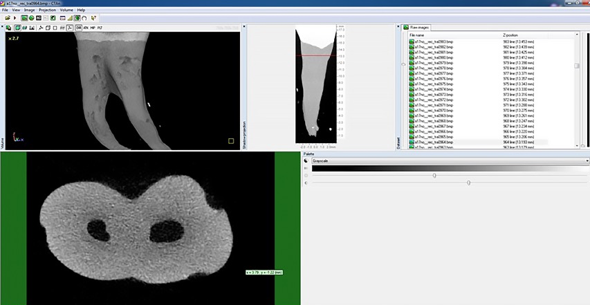
Methodology: A total of 12 maxillary first molar teeth affected by MIH were obtained from 10 children aged from 10 to 12 years. The frequency of the presence of accessory canals was examined by using microcomputed tomography and 3D image software.
Results: Accessory canals were observed in the mesiobuccal (MB) canal in all of the samples with a statistically significant difference.(p<0.05) It was observed that the accessory canals were mostly in communication with the canals in the MB root and that furcal accessory canals were found in 10 (83.33%) teeth. The incidence of accessory canals was 75% in the distobuccal (DB) canal and it was 66.66% in the palatal (P) canal.
Conclusion: The incidence of the presence of accessory canals in DB and P canals and furcation is higher in the teeth affected by MIH.
How to cite this article: Özükoç C. Examination of root canal morphology of teeth affected by Molar Incisor Hypomineralization (MIH): Frequency of accessory canals. Int Dent Res 2021;11(1):12-5. https://doi.org/10.5577/intdentres.2021.vol11.no1.3
Linguistic Revision: The English in this manuscript has been checked by at least two professional editors, both native speakers of English.
Full text article
Authors
This is an Open Access article distributed under the terms of the Creative Commons Attribution 4.0 International License (CC BY 4.0), which permits unrestricted use, distribution, and reproduction in any medium, provided the original work is properly cited.


