One-year clinical evaluation of bone-level and tissue-level implants

Article Sidebar
-
Bone-level implant, tissue-level implant, implant survival rate, mandible, tooth loss
Abstract
Aim: This study aimed to retrospectively examine and compare the clinical conditions of different implant levels at the end of 1 year. The clinical success is closely related to the loading time, the primary stability, oral hygiene, the features of the implant neck, and the bone structure.
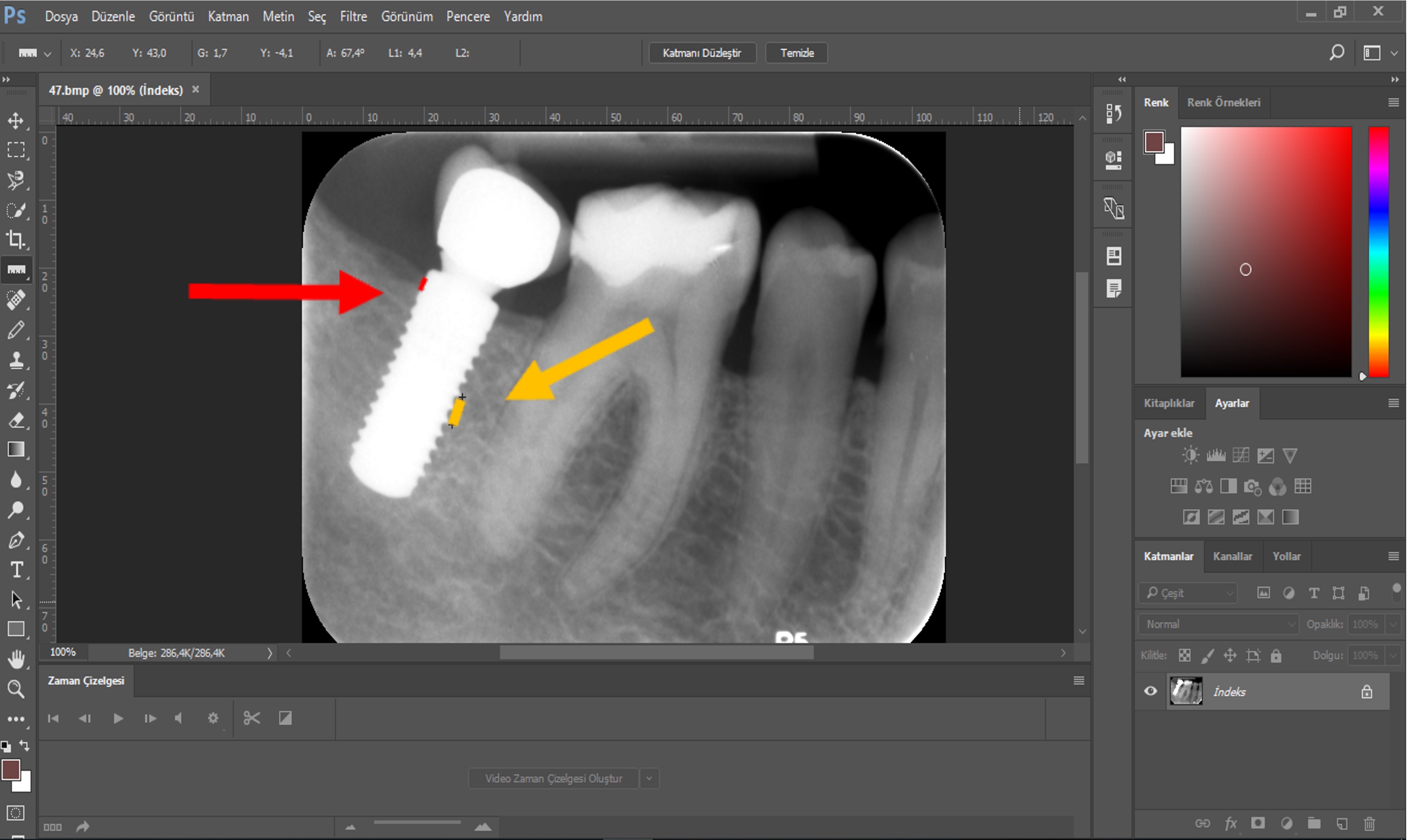
Methodology: A total of 41 implants were applied to patients with a mean age of 52.76±11.39 years. Among these patients, 10 (47.6%) were female, and 11 (52.4%) were male. Out of the total of 19 implants, 46.3% were placed in the left mandible. Specifically, 14.6% were placed in the premolar area and 31.7% were placed in the molar region. The 22 (53.7%) remaining cases were placed in the right mandible, with 10 (24.4%) in the premolar region and 12 (29.3%) in the molar region. In all, 21 implants (51.2%) were placed at the bone level, whereas 20 implants (48.8%) were placed at the tissue level. Out of the total of 41 implants, 25 (61%) had a diameter of 3.3 mm, whereas 16 (39%) had a diameter of 4.1 mm. X-rays were taken to detect bone loss in the mesial and distal regions, transferred to the computer, and evaluated via IBM SPSS Statistics V22 software.
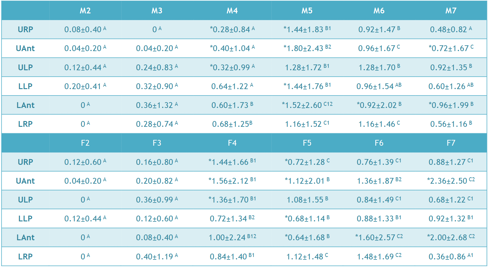
Results: In the study, 19 (46.3%) of the implants were placed in the left mandible and 22 (53.7%) in the right mandible. Approximately 51.2% of the implants were placed at the bone level, and 48.8% were at the tissue level. Initial Osstell values ranged from 72.88 ± 6.4 ISQ to 75.83 ± 5.92 ISQ. The initial X-ray results varied between 0.03 ± 0.09 mm and 0.38 ± 0.99 mm, whereas the final X-ray measurements were 0.15 ± 0.24 mm to 0.71 ± 1.54 mm.
Conclusion: Bone loss was determined to be less than 2 mm in both groups. Plaque accumulation occurred in the lingual region; however, it was observed more at the bone level. Hygiene control is the key for success, which could easily be accomplished with the gingival level of implants or the gingival positioning height of the abutments.
Full text article
Authors
Copyright © 2024 International Dental Research

This work is licensed under a Creative Commons Attribution 4.0 International License.
![]()
This is an Open Access article distributed under the terms of the Creative Commons Attribution 4.0 International License (CC BY 4.0), which permits unrestricted use, distribution, and reproduction in any medium, provided the original work is properly cited.