Open Access
78 Items
All Items
-
Examination of stress distributions on Class V restorations using nanohybrid resin composite: A 3D finite element stress analysis
Aim: The aim of this study was to investigate and compare the distribution of stress zones in composite resins and dental tissues applied to different-sized Class V cavities in the cervical region of the tooth as a result of the forces acting on the tooth in a computer environment using the finite element method. Methodology: The extracted lower first molar tooth was scanned and recorded with a dental tomography device. Images were obtained as DICOM files, and these files were imported into the Mimics 12.00 program. In this program, a 3D image of the tooth was created and converted to Standard Tessellation Language (STL) files. The obtained STL files were transferred to the Geomagic Design X program, and Standard for the Exchange of Product Model Data (STP) files were created. The SolidWorks program was used to create different sizes of Class V cavity preparation, restorative material layers, adhesive material layers, and periodontal tissues. Finally, a finite element (FE) model was created in the ABAQUS program, and stress distributions were analyzed. Results: When the depth was kept constant and the width was increased, the amount of stress on the enamel tissue decreased, while the amount of stress on all other tissues and the restorative material increased. When the width was kept constant and the depth was increased, the amount of stress on the restorative material, adhesive layer, and cortical bone increased, while the amount of stress on the enamel layer, dentin layer, periodontal ligament, and trabecular bone decreased. Conclusion: Based on the values found in the study, the model that produced the least stress on the restorative material was the model with the least depth and width. However, the stress accumulation in the enamel tissue was higher in this model compared to the other models. As a result, the lifespan of restorative materials may be longer in cavities prepared more shallowly and narrowly during the preparation phase. How to cite this article: Harmankaya A, Gönder HY, Mohammadi R, Karaköy H, Tunçdemir MT, Yüksel İB, Göktolga EG. Examination of stress distributions on Class V restorations using nanohybrid resin composite: A 3D finite element stress analysis. Int Dent Res. 2024;14(S1):90-97. https://doi.org/10.5577/indentres.562Finite element analysis, nanohybrid, composite, Class V, Von Mises stressAbdulkadir Harmankaya, Hakan Yasin Gönder, Reza Mohammadi, Hilal Karaköy, Makbule Tuğba Tunçdemir, İbrahim Burak Yüksel, Emine Gülşah Göktolga90-97 -
Influence of different crown lengths on shorter implants with different lengths, diameters and designs at atrophic posterior mandible: A finite element analysis
Aim: The aim of the study was to compare the stresses caused of different crown heights on short implants with different lengths, diameters, and designs under axial and oblique loads on the cortical bone and implant system using finite element analysis. Methodology: In the atrophic posterior mandible, four different implants of different sizes and designs were placed in the first molar region. Metal supported porcelain crowns with three different heights (10 mm, 12.5 mm, and 15 mm) were designed on the computer. A total of 12 study models were created. Axial and oblique loads were applied, and Von Mises values in the implant and abutment; maximum and minimum principal stress values in the cortical bone were evaluated by the finite element analysis method. Results: Compared to axial load, significantly higher stress values were found at the implant and cortical bone in obliquely loaded models. As the length and diameter of the implant increased, the stress on the cortical bone decreased. Compared to the threaded implant, the plateau design implant caused less stress on the bone under vertical loads, while causing more stress on the oblique. Unlike the literature, it was observed that the stresses on the implant were higher in the larger diameter implant compared to the narrower. Conclusion: As a result of the stress values obtained within the limits of the analysis, when the lengths of the crowns restored on short implants were compared, it was determined that the axial forces were at an acceptable level under all conditions. How to cite this article: Mahmutoğlu Eİ, Agüloğlu S. Influence of different crown lengths on shorter implants with different lengths, diameters and designs at atrophic posterior mandible: A finite element analysis. Int Dent Res 2023;13(3)125-132. https://doi.org/10.5577/intdentres.430 Linguistic Revision: The English in this manuscript has been checked by at least two professional editors, both native speakers of English.Short implants, implant restorations, implantology, axial load, oblique load, finite element analysisElif İrem Mahmutoğlu, Süleyman Agüloğlu125-132 -
Investigation of microhardness, surface roughness, and color change of preheated composite resins
Aim: Applying heat is a method currently used to enhance the depth of polymerization in composite resin. This study aimed to evaluate the effects of heat treatment on resin hardness, color change, and surface roughness. Methodology: In this study, 64 samples (8 mm 2 × 2 mm 2 ) were produced (3M Filtek Universal, G-aenial A’CHORD, Neo Spectra and Clearfil Majesty ES-2) using a Teflon mold (n = 8). Half of the samples were preheated to 50 o C, while the other half were polymerized at room temperature. Initial color measurements were performed using a spectrophotometer, the surface roughness was assessed with a profilometer, and the microhardness was determined using a Vickers microhardness device. Subsequently, all samples were immersed in a coffee solution at 37 o C. The color changes were documented by the seventh day. The changes in color, roughness, and hardness of the materials were examined by the thirtieth day. Results: The groups that received heat treatment exhibited higher hardness values. The unheated G-aenial A’CHORD composite resin demonstrated the most significant color change by the end of the thirtieth day, while heated and unheated Clearfil Majesty ES-2 composite samples exhibited the least color change. By the end of the thirtieth day, the surface roughness of all samples increased significantly from the baseline, with the 3M Filtek Universal samples, both heated and unheated, showing the highest surface roughness. Conclusion: According to the results of this study, heat treatment of composite resins before polymerization reduces discoloration and improves hardness. Therefore, it is recommended to heat composite resins before application. How to cite this article: Çekmez MB, Karaoğlanoğlu S, Aybala Oktay E, Aydın N, Ersöz B. Investigation of microhardness, surface roughness, and color change of preheated composite resins. Int Dent Res 2024;14(1):42-49. https://doi.org/10.5577/intdentres.523Composite resin, microhardness, surface roughness, color change, preheating, polymerizationMalhun Begüm Çekmez, Serpil Karaoğlanoğlu, Elif Aybala Oktay, Numan Aydın, Bilge Ersöz42-49 -
Investigation of the effect of the application of different full ceramic materials on stress distribution to minimally invasive posterior fixed bridge restorations by finite element analysis
Aim: The aim of this study was to examine the effect of different types of all-ceramic materials used in various designs of partial posterior indirect restorations on stress distribution through finite element analysis. Methodology: Intact mandibular first premolar and first molar teeth were selected. After being soaked in saline solution for a week, 3D radiographic images were taken using cone-beam computed tomography (CBCT). Following root canal treatment, preparations were made for inlay restorations on both teeth. CBCT images were taken again after the preparations. Data from CBCT scans were used to create 3D models in the Mimics program. Restoration designs were prepared in Standard Tessellation Language (STL) format using the EXOCAD program. The models were transferred to SolidWorks for collision processes with cement designs and then exported in Standard for the Exchange of Product Model Data (STP) format. These files were imported into the ABAQUS program for finite element analysis, with properties assigned for each restoration type. Vertical forces of 600 N were applied to the occlusal surfaces, and stress values were recorded in Von Mises Stress (VMS) values. Results: The results of the study showed that the VMS value on inlay bridge restorations was lower in mesio-occluso-distal inlay (MODI) compared to MO-DOI. ZLS exhibited the lowest VMS value for enamel loads, while nano-hybrid composite ceramics with resin matrix (RMNH) showed the lowest stress distribution in the restoration material. In posterior adhesive indirect bridge restorations, material rigidity reduced tooth load distribution. Thus, MODI preparations are preferable due to reduced stress on teeth. ZLS is advantageous as the restoration material for minimizing enamel stress. Conclusion: In cortical and trabecular bone, similar values were found across all three restorations. The highest stress concentration, excluding the area around the restoration, was observed in lithium disilicate. How to cite this article: Aydın S, Tunçdemir AR, Mohammadi R, Demirel MG. Investigation of the effect of the application of different full ceramic materials on stress distribution to minimally invasive posterior fixed bridge restorations by finite element analysis. Int Dent Res. 2024;14(S1):28-35. https://doi.org/10.5577/indentres.572Inlay bridge, indirect restorations, stress distribution, finite element analysis, Von Mises stressSalih Aydın, Ali Rıza Tunçdemir, Reza Mohammadi, Mehmet Gökberkkaan Demirel28-35 -
Investigation of the effects of various restorative materials on stress distribution in a zirconia abutment in the anterior region: A finite element analysis
Aim: The aim of this study was to investigate the effect of using aesthetic restorations and abutments in cases where implants will be applied in the anterior region on the stress distribution on implant components and surrounding biological tissues. Methodology: The data was created in the SolidWorks program, with cortical bone approximately 2 mm thick and trabecular bone approximately 15 mm thick. An implant obtained from a dental implant company, with a length of 12 mm and a diameter of 3.7 mm, was placed into the created data. The obtained data was exported as the Standard Tessellation Language (STL) file and imported into the EXOCAD program. A custom abutment and a prosthetic restoration with central incisor morphology were designed within the data, and all data were exported in STL format and re-imported into SolidWorks. Cement data with a thickness of 30 μm was created, and then all parts were assembled appropriately. The assembled data was converted to Standard for the Exchange of Product Model Data (STP) format and transferred to the ABAQUS program. In ABAQUS, zirconia was assigned to the custom abutments, dual-cure resin cement to the cement, and three different all-ceramic materials, MZ, zirconia-reinforced lithium silicate (ZLS), and lithium disilicate glass ceramics (LS), to the restoration material. A load of 150 N was applied obliquely at a 30º angle to the palatoincisal corner, and the resulting values were recorded as von Mises stress (VMS) values. Results: In each of the three restorations, the highest von Mises stress value occurred around the abutment, while the lowest stress value was observed in the trabecular bone. The stress value around the restoration was highest in monolithic zirconia. The highest stress value around the abutment was observed in lithium disilicate. Conclusion: In cortical and trabecular bone, similar values were found across all three restorations. The highest stress concentration, excluding the area around the restoration, was observed in lithium disilicate. How to cite this article: Avcı FN, Mohammadi R, Keçeci M, Demirel MG. Investigation of the effects of various restorative materials on stress distribution in a zirconia abutment in the anterior region: A finite element analysis. Int Dent Res. 2024;14(S1):106-113. https://doi.org/10.5577/indentres.569Finite element analysis, monolithic zirconia, lithium disilicate, Von Mises stress analysis, zirconia-reinforced lithium disilicate, stress distributionFatma Nur Avcı, Reza Mohammadi, Murat Keçeci, Mehmet Gökberkkaan Demirel106-114 -
Irrigation activation methods in endodontics: A comprehensive review
In contemporary endodontics, the core objective is to thoroughly cleanse and disinfect the root canal system. While shaping the canals is an important step, it alone is not sufficient. Successful treatment relies on a combined chemo-mechanical approach, which ensures the removal of organic tissue remnants, microbial contaminants, and their by-products from within the canal space. In this context, flushing the root canals with appropriate irrigation solutions is of great importance. The irrigation solutions used play a critical role in dissolving both organic and inorganic tissues and reducing the microbial load. However, the effect of irrigation can be further enhanced by actively circulating and agitating the solution within the canals. For this reason, activation methods such as ultrasonic or sonic activation systems, negative pressure irrigation techniques, and laser-assisted methods have become widely used in treatment protocols in recent years. In short, successful endodontic treatment is not only achieved by shaping the canal; it is also possible through the correct selection of irrigation solutions, sufficient duration of use, and effective activation. Once these procedures are properly carried out, the root canal system becomes more effectively disinfected, which in turn enhances the likelihood of achieving a successful long-term treatment outcome. This article comprehensively evaluates the activation devices used in today's endodontic practices and the new systems recently introduced to the market in terms of their advantages and limitations. Furthermore, current activation techniques are discussed comparatively, and their contributions to clinical practice and differences in use are debated.Endodontic, irrigation activation, sonic irrigation, passive ultrasonic irrigation, EndoActivator, EasyClean, Ultra XMakbule Taşyürek, Özkan Adıgüzel92-115 -
Knowledge, attitudes, and clinical practices of Turkish medical and dental practitioners regarding botulinum toxin use: A cross-sectional survey
Aim: This study aimed to evaluate the knowledge, attitudes, clinical practices, preferred indications, and adverse effects associated with botulinum toxin (BTX) use among medical and dental practitioners in Türkiye. Methodology: An online, self-administered questionnaire adapted from previously published studies was conducted between October 9 and December 9, 2023. A total of 276 physicians and dentists across Türkiye voluntarily participated. Participants were recruited using convenience sampling via online distribution. The survey consisted of 16 questions assessing demographic characteristics, BTX experience, indications for use, number of patients treated annually, preferred commercial formulations, adverse effects, and bruxism management preferences. Statistical analyses were performed using descriptive statistics, chi-square test, and the Fisher–Freeman–Halton exact test, with significance determined at p < 0.05. Results: A total of 53.3% of practitioners reported using BTX in clinical practice. The most common indications were bruxism (75.5%), facial wrinkles (67.3%), masseter hypertrophy (51.7%), and gummy smile (49%). OnabotulinumtoxinA was the most preferred formulation (73.5%). The most frequently reported treatment-related adverse effects were insufficient therapeutic response (49%) and localized stinging or burning pain during injection (22.4%). The injection-related adverse effects included pain (57.1%), bruising (49%), and headache (24.5%). Significant differences were observed across specialties and experience levels. BTX use was also significantly higher among practitioners working in private settings. Conclusion: The findings show that BTX was used by a considerable proportion of medical and dental practitioners in the study sample across both aesthetic and therapeutic indications. Differences observed according to workplace setting, certification status, specialty, and experience suggest that BTX-related practice patterns may not be uniform. These results highlight the potential need for structured education, certification, and standardization regarding BTX use in clinical practice.Botulinum toxin, bruxism, temporomandibular disorders, medical practitioners, dental practitioners, onabotulinumtoxinA, incobotulinumtoxinA, abobotulinumtoxinAEsra Bilgi Özyetim, Merve Dedee260695 -
Laser systems and current applications in endodontics: A review
Laser technology has gained increasing attention in endodontics due to its potential to enhance disinfection, improve treatment outcomes, and support minimally invasive clinical approaches. The aim of this review was to provide a comprehensive overview of commonly used laser systems in endodontics and to summarize their current clinical applications. Various laser types, including carbon dioxide (CO₂), diode, neodymium-doped yttrium aluminum garnet (Nd:YAG), neodymium-doped yttrium aluminum perovskite (Nd:YAP), erbium-doped yttrium aluminum garnet (Er:YAG), and erbium chromium-doped yttrium scandium gallium garnet (Er,Cr:YSGG), have been evaluated in terms of their mechanisms of action and clinical effectiveness. Lasers are used in multiple endodontic procedures, such as root canal disinfection, smear layer removal, pulp capping, pulpotomy, dentin hypersensitivity management, and treatment of pulp and periapical pathologies. In particular, laser-activated irrigation techniques, including photon-induced photoacoustic streaming (PIPS) and shock wave–enhanced emission photoacoustic streaming (SWEEPS), have shown promising results in improving irrigation efficacy and bacterial elimination, especially in complex root canal anatomies. Compared with conventional irrigation methods, these advanced laser techniques enhance the penetration of irrigants into dentinal tubules while minimizing thermal damage to surrounding tissues. Erbium lasers, owing to their high water absorption and photoacoustic effects, appear to be especially effective for smear layer removal and biofilm disruption. Other laser systems, such as diode and Nd:YAG lasers, also contribute to antimicrobial activity and postoperative pain reduction when used appropriately. However, laser parameters, irradiation time, and safety considerations remain critical for successful clinical outcomes. In conclusion, laser systems offer significant advantages in endodontic therapy by improving disinfection, supporting tissue preservation, and enabling minimally invasive techniques. Further well-designed clinical studies are needed to strengthen the evidence regarding their long-term efficacy, safety, and optimal clinical protocols.Laser, endodontics, CO₂, Nd:YAG, Nd:YAP, Er:YAG, Er,Cr:YSGG, PIPS, SWEEPSMakbule Taşyürek, Özkan ADIGÜZEL116-122 -
One-year clinical evaluation of bone-level and tissue-level implants
Aim: This study aimed to retrospectively examine and compare the clinical conditions of different implant levels at the end of 1 year. The clinical success is closely related to the loading time, the primary stability, oral hygiene, the features of the implant neck, and the bone structure. Methodology: A total of 41 implants were applied to patients with a mean age of 52.76±11.39 years. Among these patients, 10 (47.6%) were female, and 11 (52.4%) were male. Out of the total of 19 implants, 46.3% were placed in the left mandible. Specifically, 14.6% were placed in the premolar area and 31.7% were placed in the molar region. The 22 (53.7%) remaining cases were placed in the right mandible, with 10 (24.4%) in the premolar region and 12 (29.3%) in the molar region. In all, 21 implants (51.2%) were placed at the bone level, whereas 20 implants (48.8%) were placed at the tissue level. Out of the total of 41 implants, 25 (61%) had a diameter of 3.3 mm, whereas 16 (39%) had a diameter of 4.1 mm. X-rays were taken to detect bone loss in the mesial and distal regions, transferred to the computer, and evaluated via IBM SPSS Statistics V22 software. Results: In the study, 19 (46.3%) of the implants were placed in the left mandible and 22 (53.7%) in the right mandible. Approximately 51.2% of the implants were placed at the bone level, and 48.8% were at the tissue level. Initial Osstell values ranged from 72.88 ± 6.4 ISQ to 75.83 ± 5.92 ISQ. The initial X-ray results varied between 0.03 ± 0.09 mm and 0.38 ± 0.99 mm, whereas the final X-ray measurements were 0.15 ± 0.24 mm to 0.71 ± 1.54 mm. Conclusion: Bone loss was determined to be less than 2 mm in both groups. Plaque accumulation occurred in the lingual region; however, it was observed more at the bone level. Hygiene control is the key for success, which could easily be accomplished with the gingival level of implants or the gingival positioning height of the abutments. How to cite this article: Sözkes S, Kayabay G, Koçak Büyükdere A. One-year clinical evaluation of bone-level and tissue-level implants. Int Dent Res 2024;14(2):50-56. https://doi.org/10.5577/idr2024.392Bone-level implant, tissue-level implant, implant survival rate, mandible, tooth lossSarkis Sözkes, Gökçe Kayabay, Ayse Koçak Büyükdere50-56 -
Ozone applications in dentistry: A review
Ozone (O 3 ) therapy has been applied in medicine and dentistry for many years and has become increasingly widespread. Ozone, composed of three oxygen atoms (O 3 ), is a strong antioxidant and a highly effective antimicrobial agent. Ozone gas has high oxidizing properties and exhibits a 1.5-fold higher antimicrobial effect than chloride. Its use has become popular in many fields of dentistry in recent years, owing to its various properties. It is categorized as one of the non-invasive, atraumatic, protective, and preventive treatment modalities that have been improved by modern dentistry, and its acceptability among patients has increased as a result. Ozone is an antimicrobial, analgesic, and biosynthetic agent that stimulates the immune system, enhances blood circulation capacity, and positively affects oxygen transport mechanisms. It influences both cellular and humoral immunity. All three forms of ozone (gaseous, oil, and solution) can be used in dentistry. It is indicated for the treatment of approximately 300 different pathological conditions. Ozone is used in endodontics (irrigation solution, intracanal medicament, abscess cases, disinfection), periodontology (gingivitis, periodontitis, periimplantitis, surgical injuries, prophylaxis), oral pathology (stomatitis, aphthous ulceration, candidiasis, herpes lesions), oral, dental, and jaw surgery (implantation, reimplantation, simple-to-complicated tooth extraction, homeostasis, wound healing, temporomandibular joint (TMJ) surgery, osteonecrosis cases), prosthetic dental treatment (disinfection of fixed and removable prostheses), restorative dental treatment (caries lesions, dentine hypersensitivity, cracked tooth, bleaching), orthodontics (remineralization and prophylaxis of white spot lesions), pedodontics (caries lesions, root canal treatment, positive psychological impact, short treatment duration), and sterilization of dental instruments and units. This review aimed to describe the usage fields and effects of ozone in dental applications, provide insights into clinical practices in other fields of dentistry, and introduce newly conducted in vitro and in vivo studies.Ozone, ozone therapy, dentistry, disinfectionMerve Doğan İlik, Özkan Adıgüzel, Myroslav Goncharuk-Khomyn222-236 -
Performance of ChatGPT-5, Gemini 2.5 Flash, and DeepSeek-V3.2 large language models in response to questions about ozone in dentistry
Aim: The aim of this study was to compare and evaluate the accuracy and completeness of responses to open-ended questions about the use of ozone in dentistry that were addressed to three recent large language models (LLMs): ChatGPT-5, Gemini 2.5 Flash, and DeepSeek-V3.2. Methodology: A total of 25 open-ended questions on the subject of ozone in dentistry were asked to three different LLMs (ChatGPT-5, Gemini 2.5 Flash, and DeepSeek-V3.2). The chat history was deleted after each response, and a new session was started for each question. All questions were asked only once using the same computer IP address, on the same day, on the same fixed fiber Internet network. The questions were asked to the chatbots in English, and English responses were obtained and recorded. The responses were evaluated independently by two restorative dentistry specialists using two separate Likert scales for accuracy and completeness. In the statistical analyses, the Intraclass Correlation Coefficient (ICC) was calculated, and the Shapiro–Wilk, Kruskal–Wallis, Dunn–Bonferroni, and Spearman tests were applied. Results: The agreement between the evaluators was found to be high in respect of the accuracy points and varied according to the model for the completeness points. Significant differences were determined between the LLMs in the comparisons of the accuracy scores (p < 0.001). The median points of the ChatGPT-5 and DeepSeek-V3.2 models were determined to be statistically significantly higher than those of Gemini 2.5 Flash (p = 0.002 and p < 0.001, respectively). Significant differences were observed between the LLMs in terms of completeness scores (p = 0.004). In the subgroup analyses, the mean points of the ChatGPT-5 and DeepSeek-V3.2 models were determined to be statistically significantly higher than those of Gemini 2.5 Flash (p = 0.006 and p = 0.021, respectively). Conclusion: The responses to questions about the use of ozone in dentistry showed differences among the LLMs in terms of accuracy and completeness. ChatGPT-5 and DeepSeek-V3.2 demonstrated better performance than Gemini 2.5 Flash. Despite the high accuracy rates, it must not be forgotten that incorrect healthcare information can lead to serious outcomes. Therefore, it should be recommended that LLMs be used under human supervision in clinical applications and that future research focus on increasing the diagnostic reliability of artificial intelligence.Artificial intelligence, large language models, ChatGPT, Gemini, DeepSeek, ozone, dentistrySuzan Cangül, Özkan Adıgüzel, Makbule Taşyürek, Tuba Tunç, Hatice Ortaç165-174 -
Performance of two different electronic apex locators during the removal of calcium silicate-based sealers
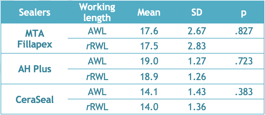
Aim: The aim of this in vitro study is to evaluate the accuracy of Root ZX and Propex Pixi during the retreatment of root canals obturated with different calcium silicate-based root canal sealers. Methodology: Root canals of ninety human premolar teeth were shaped with ProTaper universal rotary files up to size F2 under 5.25% NaOCl irrigation for the present study. Root canal filling was carried out by lateral condensation technique using with gutta-percha and sealers; MTA Fillapex, CeraSeal or AH Plus. After seven days, the obturating materials were removed using D-Race files. While actual working length was recorded by direct observation, electronic working lengths were determined by Propex Pixi or Root ZX for tolerance limits of 0.5 mm and 1.0 mm. Data were analyzed using Shapiro Wilk, Mann-Whitney U, and Kruskal-Wallis tests ( p < 0.05). Results: The performance of Propex Pixi and Root ZX was found to be statistically similar and did not differ on the root canals filled with different root canal sealers ( p < 0.05). Propex Pixi and Root ZX demonstrated more accurate measurements in the range of ±1 mm compared to ±0.5 mm in all groups, regardless of the type of root canal sealer used when filling the root canals before retreatment ( p < 0.05). Conclusion: Root ZX and Propex Pixi can be used confidently in the retreatment of the root canals in which CeraSeal, MTA Fillapex, and AH Plus are used as sealers. How to cite this article: Göçer PA, Durmazpınar PM, Baştürk FB. Performance of two different electronic apex locators during the removal of calcium silicate-based sealers. Int Dent Res 2023;13(S1):20-25. https://doi.org/10.5577/idr.2023.vol13.s1.3 Linguistic Revision: The English in this manuscript has been checked by at least two professional editors, both native speakers of English.Apex locator, apical constriction, calcium silicate-based sealer, retreatment, working lengthPelin Andaç Göçer, Parla Meva Durmazpınar, Fatima Betül Baştürk20-25 -
Prevalence of radix entomolaris and radix paramolaris in mandibular first and second molars in a Turkish population
Aim: This retrospective study determined the prevalence and distribution of extra roots (radix entomolaris [RE] and radix paramolaris [RP]) in mandibular first and second molars in a southeastern Anatolian Turkish population using cone-beam computed tomography (CBCT). Methodology: A total of 2010 mandibular first and second molars from the archived CBCT images of 662 patients (325 males, 337 females) were evaluated for the associations of RE and RP with sex, side, molar type, and unilateral/bilateral presence. Fisher's exact test was used for categorical comparisons and McNemar's exact test for the paired right-left comparison at patient level; prevalences are reported with Wilson score 95% confidence intervals, and the 24 pairwise post-hoc comparisons were corrected for multiplicity by the Holm procedure. Results: An extra root was detected in 3.7% of the examined teeth (75/2010, 95% CI 3.0-4.7%) and 9.5% of the patients (63/662, 95% CI 7.5-12.0%). Prevalence did not differ significantly by sex (8.9% [29/325] in males versus 10.1% [34/337] in females, p = 0.61), side (McNemar exact p = 0.16), or unilateral/bilateral presence (p = 1.00). Although the overall per-tooth prevalence was similar in first and second molars (3.9% versus 3.6%, p = 0.73), the distribution of root type among the 75 affected teeth differed significantly (Fisher exact p = 0.002). RP was the most frequent type in both first (1.95%, 19/974) and second (3.09%, 32/1036) molars but exceeded RE significantly only in the second molars (RE 4/1036, p < 0.001; first molars RE 14/974, p = 0.49). Among the affected teeth, RP was relatively more common in the second molars (Holm-adjusted p = 0.025), whereas the excess of RE in the first molars did not survive correction for multiple testing (Holm-adjusted p = 0.26). Conclusion: The presence of extra roots in mandibular molars within the Turkish population is a key variation for access cavity preparation in endodontics. The different distributions of these roots between the first and second molars emphasize the need for a thorough pre-treatment radiographic assessment.Cone-beam computed tomography, root canal anatomy, prevalence, tooth abnormalities, radix entomolaris, radix paramolarisAyşenur Çam, Merve Defişet, Merve Yeniçeri Özata, Özkan Adıgüzele260663 -
Psychosocial impact of COVID-19 among adolescent orthodontic patients: A multicenter cross-sectional study
Aim: The aim of this study was to assess the COVID-19 anxiety and fear levels of adolescent patients undergoing orthodontic treatment in terms of their demographic variables, treatment location, COVID-19 status, and various clinical conditions (hospitalization and psychological support), as well as to evaluate their associations with age and treatment duration. Methodology: A multicenter descriptive and cross-sectional study was conducted in four different private clinics and a public hospital during the pandemic. Descriptive data included information on demographics, orthodontic treatment, and COVID-19 status. Anxiety and fear were assessed using the COVID-19 Anxiety Scale (CAS) and Fear of COVID-19 Scale (FC-19S), respectively. The collected data was statistically analyzed with the Shapiro-Wilk, Student’s t, Mann-Whitney U, and Kruskal-Wallis (Dunn post hoc) tests ( p <0.05). Results: A total of 231 adolescent patients (133 female and 98 male) with a mean age of 15.06 ± 1.61 years were examined. Higher anxiety scores were observed in patients from high-income families. In addition, patients receiving orthodontic treatment in private clinics reported significantly higher anxiety scores ( p <0.05). Patients were more anxious and experienced fear when they had COVID-19-related symptoms or were exposed to COVID-19-positive patients ( p <0.05). A positive correlation was found between the CAS and FC-19S scores (r =0.425; p =0.001) and between the treatment duration and FC-19S scores (r = 0.252; p =0.025). Conclusion: In the presence of COVID-19-like pandemic disease, private clinicians should be aware of delayed appointments to reduce the anxiety level of their patients, taking into account their income level and exposure to infection.adolescent, anxiety, COVID-19, fear, anxiety scale, fear scaleEyüp Burak Küçük, Serpil Çokakoğlu, Mehmet Hanifi Kokaçya, Mehmet Karadağ30-37 -
Regenerative endodontics: A review
Oral health and function are critical components of a patient's overall quality of life. Pulp necrosis in immature permanent teeth, resulting from trauma, caries, or developmental anomalies, presents a significant clinical challenge. Traditionally, apexification has been the routine treatment; however, it fails to promote continued root development, often leaving teeth brittle and susceptible to fractures. This review examines regenerative endodontic procedures (REPs), which represent a paradigm shift toward biologically based treatments designed to replace damaged structures and cells of the pulp-dentin complex. The success of REPs relies on the interaction of three key components: stem cells, bioactive molecules (growth factors), and scaffolds. During these procedures, a blood clot is typically induced to act as a scaffold, facilitating the influx of undifferentiated stem cells, such as stem cells of the apical papilla (SCAPs). The review details current clinical protocols recommended by the American Association of Endodontists (AAE) and the European Society of Endodontology (ESE). Key technical aspects include the use of low-concentration sodium hypochlorite (1.5%) to maintain cell viability and EDTA to promote the release of growth factors from the dentin matrix. Intracanal medicaments, specifically triple antibiotic paste (TAP) and calcium hydroxide, are evaluated for their disinfection efficacy and impact on stem cell survival. While REPs demonstrate high success rates in terms of symptom resolution and apical healing, histological evidence suggests the resulting tissue is often a repair (bone or cementum-like) rather than true pulpal regeneration. Despite these challenges, REPs offer a promising therapeutic pathway for increasing root length and wall thickness, ultimately enhancing the long-term retention of immature permanent teeth.Regenerative endodontics, pulp revitalization, immature permanent teeth, Stem Cells of the Apical Papilla, SCAP, tissue engineering, triple antibiotic paste, root development, scaffoldsTolga Han Edebal, Gülşen Edebal203-221 -
Role of CBCT and non-surgical endodontic management of misdiagnosed odontogenic extraoral sinus tracts: Three case reports
Odontogenic extraoral sinus tracts are uncommon clinical conditions that frequently present diagnostic challenges because of their atypical extraoral appearance and the frequent absence of dental symptoms. Consequently, these lesions are often misdiagnosed as dermatologic or systemic diseases, resulting in inappropriate treatments, prolonged morbidity, and delayed healing. Accurate identification of the odontogenic source is essential for definitive management. These case reports describe three female patients who presented with persistent extraoral sinus tracts of odontogenic origin that had been previously misdiagnosed and managed as dermatologic lesions. All patients had undergone prolonged topical or conservative treatments without clinical improvement. Comprehensive clinical examination and conventional radiography, supplemented by cone-beam computed tomography (CBCT), revealed necrotic pulps associated with periapical lesions, accessory root canals, and localized bone defects in the affected teeth. All cases were treated using a non-surgical endodontic approach. CBCT played a critical role in detecting complex root canal anatomy and guiding treatment planning. Root canal disinfection protocols included irrigation with sodium hypochlorite and ethylenediaminetetraacetic acid, ultrasonic activation, and the use of calcium hydroxide as an intracanal medicament, between appointments. Final obturation was performed using gutta-percha and a bioceramic sealer, followed by definitive coronal restoration. Clinical follow-up demonstrated progressive resolution and complete closure of the extraoral sinus tracts in all patients. At the one-year follow-up, complete soft tissue healing and radiographic evidence of periapical bone regeneration were observed, with no signs of recurrence. All treated teeth remained asymptomatic and functional during the follow-up period. These cases emphasize the importance of including odontogenic origins in the differential diagnosis of persistent extraoral facial lesions. CBCT is a valuable diagnostic adjunct in complex cases, particularly for identifying accessory canals and periapical pathologies. When accurately diagnosed, non-surgical endodontic treatment provides a predictable, minimally invasive alternative to surgical intervention, enabling complete functional and esthetic healing while preventing unnecessary dermatologic or surgical procedures.Cone-beam computed tomography, odontogenic infections, sinus tract, root canal treatment, bioceramic sealerCerengül Çandır, Neslihan Şimşek, Elçin Tekin Bulut -
Stress distribution of customized abutment material and angles in dental implant systems: A finite element study
Aim: This study aims to use finite element analysis to evaluate stress distribution in implant components, restoration materials, and biological tissues for implants placed at different angles using various customized abutment materials. Methodology: 3D models of a three-member implant-supported fixed prosthesis were created using SolidWorks, with implants placed at positions 44 (4I) and 46 (6I). These models were imported into EXOCAD to design prosthetic restorations on custom abutments at three angles: straight (KD0), 10° (KD10), and 20° (KD20). Abutments were made from alumina (AL), titanium (TI), and zirconia (ZI). A 150 N load was applied to the occlusal table at a 30° angle in the buccolingual direction. Stress distribution was measured as von Mises Stress (vMS) values. Results: For implants, the lowest vMS value was observed in the KD0-AL group (4I: 9.984 MPa, 6I: 22.91 MPa), while the highest vMS value was found in the KD20-TI group (4I: 25.30 MPa, 6I: 42.03 MPa). Custom abutment and custom abutment screws (in general) showed higher vMS values in the AL material and lower vMS values in the TI material. In contrast, for cortical bone and restoration material, higher vMS values were observed in the TI material and lower vMS values in the AL material. Conclusion: Dental implants require effective stress distribution from occlusal loads to ensure long-term success. Excessive stress on the abutment and abutment screw can compromise the prosthetic outcome. Therefore, titanium (TI) abutments are preferred in the posterior region for their durability despite being less aesthetically pleasing. Additionally, a higher implant angle reduces strength, negatively impacting the implant's performance. How to cite this article: Mohammad R, Demirel MG, Erdoğdu M. Stress distribution of customized abutment material and angles in dental implant systems: A finite element study. Int Dent Res 2024;14(S1):1-11. https://doi.org/10.5577/indentres.567Custom abutment, finite element analysis, implant-supported prostheses, stress distribution, Von Mises stressReza Mohammadi, Mehmet Gökberkkaan Demirel, Meryem Erdoğdu1-11 -
Support vector machine-based diagnostic classification of caries and healthy teeth in panoramic orthopantomography images
Aim: This study proposes a support vector machine (SVM)-based classification model for detecting dental caries in panoramic dental orthopantomography (OPG) images. The primary objective is to develop a computationally efficient, reproducible, and clinically adaptable method that enables the automatic differentiation between carious and healthy tooth structures. Methodology: The images used in the study were first subjected to standard resizing and normalization steps. The extracted features were then prepared for classification through feature scaling, feature selection, and dimensionality reduction processes. Hyperparameter grid search, stratified k-fold cross-validation, and decision threshold optimization were applied during the model development process. Medically meaningful metrics, including accuracy, precision, sensitivity, F1 score, area under the receiver operating characteristic (ROC) curve (AUC), and the confusion matrix, were used in the performance evaluation. Results: The SVM model demonstrated balanced and stable performance throughout cross-validation. ROC and Precision–Recall analyses showed that the model has a high capacity to distinguish between decayed and healthy classes. Decision threshold scanning revealed that false-positive and false-negative rates can be optimized according to clinical requirements. Conclusion: The results show that the proposed SVM-based approach offers a reliable and feasible method for automatic caries detection from panoramic OPG images. This study highlights that traditional machine learning methods, when combined with structural feature engineering, are suitable for supporting dental diagnosis processes. Future studies should test the model on larger datasets and perform multi-classification of different dental pathologies.Support vector machine, machine learning, deep learning, hyperparameter, dentistry, panoramic dental radiography, caries classificationSüleyman Dal, Yasin Sönmez136-147 -
The application of ultrasonography to the masticatory system: A bibliometric analysis
Aim: This study aimed to summarize the current knowledge on ultrasound use in the masticatory system and establish a scientific foundation for future research. Methodology: A literature search of the Web of Science database was completed in August 2024, following preliminary scans and searches conducted before August 2, 2024, to refine the approach. A total of 205 studies were included in the meta-analysis. Bibliometric analysis and data visualization were performed using VOSviewer and the R package Bibliometrix (Biblioshiny). Results: The first study on this topic was published in 1991. The highest number of publications and citations occurred in 2023. Turkey was the top producer of articles, whereas Japan had the greatest impact. The Universidade Estadual de Campinas published the most papers, and the Academic Center for Dentistry, Amsterdam, was the most cited institution. Stavros Kiliaridis emerged as the most prolific author, and Oral Surgery, Oral Medicine, Oral Pathology and Oral Radiology (OOOO Journal) was the most productive journal. Conclusion: This study conducted a comprehensive bibliometric analysis and visualization of research on ultrasound use in the masticatory system using literature from the Web of Science Core Collection database covering 1991 to 2024. The current focal areas and future research avenues were identified. This research also clarified the historical foundations and prevailing trends in ultrasound application within this field, offering a nuanced perspective for scholars.bibliometrics, masseter muscle, masticatory system, ultrasonographyİbrahim Tevfik Gülşen, Ruşen Erdem, Yavuz Selim Genç, Emine Gülşen18-29 -
The effect of different implant-bone levels on stress distribution in three-unit monolithic zirconia restorations: A 3D finite element analysis
Aim: The aim of this study is to investigate the stress distribution in implant-supported three-unit monolithic zirconia restorations according to different bone levels at which implants are placed in the mandible, using three-dimensional finite element analysis. Methodology: For this study, mucosal, trabecular, and cortical bone pieces were designed using the SolidWorks program to simulate an edentulous mandibular jawbone. Two implants were positioned in areas numbered 44 and 46 with different subcrestal placements (0 mm, 0.5 mm, 1.0 mm, 1.5 mm, 2.0 mm). In each model, dental implants, abutments (TiBase), monolithic zirconia crowns, abutment screws, and bone tissues were simulated. The obtained data were exported as Standard Tesselation Language (STL) files and imported into the Exocad program for the design of prosthetic restorations and custom abutments. The STL files were then imported into the Geomagic Design X program to merge the implants, abutments, and restorations on the digital model. The final models were saved as STP files and transferred to the ABAQUS software for finite element analysis. Subsequently, an oblique load of 150 N in the buccolingual direction was applied to the occlusal table for 1000 cycles, and the mandibular jawbone was fully constrained (encastre) in all directions (both movement and rotation). Stress distribution values were recorded in von Mises (vMS) units, and fatigue performances were measured using the Seegers method. Results: As the implant bone levels increased, the vMS increased on the prosthetic restoration. Specifically, vMS increased on the abutment of implant 44 but decreased on implant 46. Fatigue performance worsened on the abutment of implant 44 but improved on the abutment, and screws of implant 46. Abutment screws proved most durable, with abutments experiencing early failures. Conclusion: According to the results of the study, it has been observed that the implant bone level affects the stresses occurring in prosthetic restoration. How to cite this article: Alioğlu Koparal NS, Tunçdemir AR, Demirel MG, Mohammadi R, Keçeci M. The effect of different implant-bone levels on stress distribution in three-unit monolithic zirconia restorations: A 3D finite element analysis. Int Dent Res. 2024;14(S1):81-89 https://doi.org/10.5577/indentres.566Zirconia, implant-bone level, monolithic zirconia, finite element analysis, stress distribution, Von Mises stressNur Sena Alioğlu Koparal, Ali Rıza Tunçdemir, Mehmet Gökberkkaan Demirel, Reza Mohammadi, Murat Keçeci81-89 -
The effect of different materials and multi-unit angles in stress distribution on implant-supported fixed bridges: A finite element method study
Aim: In implant-supported restorations, various abutment angulations can be used. These angles are determined based on factors such as the structure of the jawbone, the position of the implants, and the design of the restoration. The aim of this study is to evaluate the effect of multiunit abutment angles on stress patterns of the implant and the peri-implant tissue. Methodology: In this finite element analysis study, three different three-dimensional models of maxillary 3-unit fixed implant-supported prostheses were examined. A jawbone was created with jaw components in SOLIDWORKS. Subsequently implant models obtained from an implant company(Bilimplant®) were placed in the area of the maxillary premolar, and the other implant were placed in the area of the maxillary first molar. Multiunit abutments were used at three different angles (0°, 15°, 30°). Two different restorative materials (Co-Cr feldspathic and zirconia feldspathic) were designed in EXOCAD software for the fixed implant-supported restoration. 150 newtons at a 30-degree angled oblique force was applied to the implants. Subsequently, stress distribution was evaluated with finite element analysis and performed using the ABAQUS software and analyzed with Von mises stress distribution. Results: With the increase in the multiunit angle in implant-supported prostheses, it has been observed that the stress values applied to the implant components and cortical bone increase, while the stress applied to the prosthesis decreases. The highest stress values on implant components and cortical bone were detected in the zirconia framework models. Conclusion: This study evaluates the impact of multiunit abutment angles on implant-supported restorations and examines the stress patterns of implants and peri-implant tissues. The study shows that increasing the multiunit angle in implant-supported prosthesis increases stress values on implant components and cortical bone while decreasing stress applied to the prosthesis.Implant, abutment, finite element method, angle, Von Mises stressMustafa Demirtaş, Doğan Ilgaz Kaya, Reza Mohammadi, Mehmet Gökberkkaan Demirel70-80 -
The effect of different surface treatments on the color change of CAD/CAM restorative materials caused by coffee
Aim: This study aimed to evaluate the effect of different surface treatment protocols on the color stability of two CAD/CAM restorative materials, a hybrid ceramic (Vita Enamic) and a resin-based composite (Shofu Block HC), when exposed to filter coffee. Methodology: Sixty disk-shaped specimens (12 mm diameter, 1.5 mm thickness) were prepared from Vita Enamic and Shofu Block HC. The specimens were divided into six groups (n=10) based on the material and surface treatment: Mechanical Polishing Only, Glaze (Optiglaze) Only, and Mechanical Polishing + Glaze. After the surface treatments, baseline color measurements were performed using a spectrophotometer (Vita EasyShade V). The specimens were then immersed in a freshly prepared filter coffee solution, which was renewed daily for 15 days (simulating ~3 years of clinical exposure). Post-immersion color measurements were taken, and the color difference (ΔE) and lightness difference (ΔL) were calculated. Data were analyzed using two-way ANOVA and Tukey's HSD test (p < 0.05). Results: Two-way ANOVA revealed significant main effects of Material and Surface Treatment on both ΔE and ΔL (p < 0.001). A significant Material × Surface Treatment interaction was found for ΔL (p = 0.012) but not for ΔE (p = 0.207). Vita Enamic demonstrated significantly lower ΔE and higher ΔL values than Shofu Block HC, indicating better overall color stability and less darkening. For both materials, Mechanical Polishing Only resulted in significantly lower ΔE than glaze only or the combination group. For ΔL, Mechanical Polishing was superior for Vita Enamic, whereas Glaze Only yielded the most unfavorable results for Shofu Block HC. Conclusion: The color stability of CAD/CAM materials is significantly influenced by both material composition and surface treatment applied. Mechanical polishing provided the best color stability for both materials tested. Clinically, for restorations prone to coffee exposure, selecting a hybrid ceramic like Vita Enamic and finishing it with mechanical polishing is recommended to optimize long-term aesthetic performance.CAD-CAM, color stability, dental polishing, composite resin, coffee, surface propertiesMeryem Erdoğdu, Gözde Kaya Oğuz, Merve Karagöz, Ali Rıza Tunçdemir, Emine Begüm Büyükerkmen183-194 -
The effect of immediate dentin sealing on fracture resistance and failure modes of resin composite onlay restorations
Aim: This study aimed to investigate the fracture resistance, failure modes, and reparability of molars restored with laboratory-processed resin composite (Ceramage, SHOFU Inc., Kyoto, Japan) onlays above or below the cemento-enamel junction (CEJ) in the presence or absence of immediate dentin sealing (IDS). Methodology: Forty extracted sound human molars were selected and divided into four groups: 1) Below CEJ with IDS, 2) Below CEJ without IDS, 3) Above CEJ with IDS, and 4) Above CEJ without IDS. Standardized mesio-occlusal (MO) onlay preparations were made with the reduction of mesio-buccal and mesio-palatinal cusps. Butt-joint preparation was used in the proximal box area extending 1 mm below the CEJ (Groups 1 and 2) or 1 mm above the CEJ (Groups 3 and 4). In Groups 1 and 3, IDS was applied immediately after tooth preparation. After cementation (Variolink N, Ivoclar Vivadent AG, Schaan, Liechtenstein), the specimens were thermally aged (20000 cycles, 5–55 ºC) and then subjected to load to failure (1 mm/min). The failure types and reparability of the onlays were recorded, and the data were analyzed using two-way ANOVA and Fisher’s exact tests. Results: The restorations in Group 4 showed the highest mean fracture strength compared to Group 1 ( p =0.038), Group 2 ( p =0.008), and Group 3 ( p =0.019). The interaction between the level of proximal extension and the presence of IDS did not influence fracture mode or reparability ( p >0.05). Conclusion: No difference in the fracture strength of resin composite onlays below CEJ with or without IDS was found; however, not using IDS for resin composite onlays above CEJ appears to be preferable. How to cite this article: Gözetici Çil B, Köymen SS, Ünal S, Dönmez N. The effect of immediate dentin sealing on fracture resistance and failure modes of resin composite onlay restorations. Int Dent Res 2024;14(1):27-33. https://doi.org/10.5577/intdentres.510Onlays, fracture strength, immediate dentin sealing, composite resinBurcu Gözetici Çil, Safiye Selin Köymen, Serpil Ünal, Nazmiye Dönmez27-33 -
The effect of ionizing radiation from dental radiographic devices on dental pulp stem cells
Aim: This study aims to investigate the effects of exposure to different doses delivered by radiographic diagnostic tools on cell death pathways in isolated dental pulp stem cells on a molecular level using qRT-PCR data. Methodology: Seventy fully impacted mandibular third molars were used for the study. Extracted third molars were divided into seven groups (n = 10 each): A control group receiving no radiation, periapical (PER), panoramic (PAN), and dental tomography (CBCT) groups receiving one dose of radiation each, and periapical, panoramic, and CBCT groups receiving three consecutive doses of radiation each. Then, gene expressions in dental pulp stem cells were evaluated using the qRT-PCR this method. Results: A significant difference was found in the expression of the ULK1 gene in the dental pulp tissue samples from the PAN1 group; GABARAP, LAMP3, APAF1, BCL2, CASPASE2, and CASPASE7 genes from the PER1 group; the ULK1 gene from the PER3 group; and ATG12, ATG16L1, ATG4A ATG5, BECN1L1, GABARAP, LAMP1, LAMP3, MAP1LC3B, ULK1, APAF1, BCL2, CASPASE-2, CASPASE-3, CASPASE-7, and CASPASE-8 genes from the CBCT3 group. Conclusion: While the radiation doses we applied to the DPSC cell culture are considered low levels, there was an increase in cell death markers with increased exposure to radiation through multiple radiography procedures. Taking measures to prevent errors during radiography and avoiding repeated, unnecessary patient exposure to radiation would help minimize the damaging effects of radiation. How to cite this article: Kahraman OE, Karaman T, Talo Yıldırım T, Önalan E, Dündar S, Tektemur A. The effect of ionizing radiation from dental radiographic devices on dental pulp stem cells. Int Dent Res 2023;13(3):112-8 https://doi.org/10.5577/intdentres.426 Linguistic Revision: The English in this manuscript has been checked by at least two professional editors, both native speakers of English.Dental pulp stem cells, rtPCR, mRNA expression, periapical radiography, panoramic radiography, cone-beam computed tomographyOnur Evren Kahraman, Tahir Karaman, Tuba Talo Yıldırım, Ebru Önalan, Serkan Dündar, Ahmet Tektemur112-118 -
The relationship between mandibular symphysis shape and lower incisor dentoalveolar compensation in Class I and Class III skeletal patterns: A CBCT study
Aim: The aim of this study was to examine the relationship between the lower incisor dentoalveolar compensation mechanism and the morphology of the mandibular symphysis in patients with Class I and Class III malocclusions, who have different vertical growth patterns, using cone-beam computed tomography (CBCT). Methodology: This retrospective study analyzed the CBCT scans of 60 patients, divided into four groups based on dental and skeletal characteristics: Class I normodivergent, Class I hyperdivergent, Class III normodivergent, and Class III hyperdivergent. Measurements focused on the midsagittal section of the right lower central incisor to assess the relationship between mandibular symphysis morphology and alveolar bone thickness. Results: There were no substantial differences observed in alveolar bone thickness or linear mandibular symphysis measures between the groups. However, significant differences were found in symphysis concavity, basal and dentoalveolar inclination, and Incisor Mandibular Plane Angle (IMPA) values ( p =0.036, p =0.001, p =0.003, and p =0.001, respectively). Further analysis revealed strong correlations between symphysial thickness, total symphysis height, dentoalveolar inclination, and convexity angle with mandibular symphysis-related alveolar bone thickness ( p <0.05). Conclusion: A positive correlation was observed between the dentoalveolar symphysis and alveolar bone thickness in all groups. In Class I patients, symphysis height was negatively correlated with alveolar bone thickness, whereas no correlation was found for symphysis thickness or inclination. In Class III patients, the symphysis shape provided some insight, but could not reliably estimate the alveolar bone thickness.alveolar bone thickness, Class I malocclusion, Class III malocclusion, normodivergent, hyperdivergent, cone-beam computed tomography, mandibular symphysis, symphysis inclinationRuşen Yetgin, Sabahattin Bor, Numan Dedeoğlu, Samet Özden1-9 -
The relationship between xerostomia and periodontal status in patients with cardiovascular diseases and type 2 diabetes
Aim: The aim of this cross-sectional study was to examine the correlation between patients' subjective perception of xerostomia and their periodontal status and to examine the potential association of this correlation with cardiometabolic disorders. Methodology: A total of 147 patients (40 systemically healthy, 33 with cardiovascular disease [CVD], 40 with type 2 diabetes, and 34 with cardiometabolic disease [CVD + diabetes]) who came to Necmettin Erbakan University, Faculty of Dentistry, Department of Periodontology for periodontal treatment and agreed to participate were included in the study. Sociodemographic information, tooth brushing frequency, systemic disease status, and clinical periodontal parameters (plaque index [PI], gingival index [GI], bleeding on probing [BOP], and mean pocket depths) were recorded. Patients were asked to complete an 11-item subjective xerostomia inventory questionnaire. Results: In the findings of our study, there was no statistically significant correlation between xerostomia total score and mean pocket depths, PI, GI, and BOP ( p >0.05). There was no statistically significant difference between the mean values of xerostomia total score according to systemic disease groups ( p =0.654). There is a statistically significant difference between the mean values of xerostomia total score according to periodontal status groups ( p =0.002). The mean xerostomia total score was 20.19 in periodontally healthy patients, 24.42 in gingivitis patients, and 22.73 in periodontitis patients. Conclusion: Although the results suggest that subjective dry mouth findings are associated with gingivitis, they do not affect pocket depth and do not increase susceptibility to periodontitis. Clinicians should emphasize the risk of gingivitis in patients with dry mouth and the importance of prevention. How to cite this article: Babayiğit O, Ögütcen Ö, Özkan Şen D, Taştan Eroğlu Z, Uçan Yarkaç F. The relationship between xerostomia and periodontal status in patients with cardiovascular diseases and type 2 diabetes. Int Dent Res. 2024; 14(S1):21-27. https://doi.org/10.5577/indentres.556Cardiometabolic diseases, gingivitis, periodontal status, xerostomia, type 2 diabetesOsman Babayiğit, Ömer Ögütcen, Dilek Özkan Şen, Zeynep Taştan Eroğlu, Fatma Uçan Yarkaç21-27 -
Use of novel polyetheretherketone (PEEK) polymer in pediatric dentistry
The purpose of this study is to discuss the applications of PEEK in pediatric dentistry and the advantages of PEEK material in terms of periodontal health. In pediatric dentistry, there is a continuous search for biocompatible, esthetic, and durable materials to overcome the limitations of conventional options such as stainless steel and zirconia. Polyetheretherketone (PEEK), a high-performance semicrystalline polymer, has emerged as a novel alternative due to its superior mechanical properties, resistance to heat and chemicals, and an elastic modulus comparable to human bone. This review aims to evaluate the material characteristics of PEEK, its impact on periodontal health, and its clinical applications within pediatric dentistry. PEEK is amenable to digitalization and can be fabricated using CAD/CAM milling and 3D printing, ensuring precise fit and patient comfort. In pediatric orthodontics and space maintenance, PEEK serves as an esthetic, metal-free alternative, eliminating risks of nickel allergy while providing high strength and significantly reduced weight compared to traditional appliances. For restorative procedures, PEEK is utilized in crowns, endocrowns, and post-core systems. Its shock-absorbing capability allows for favorable stress distribution in dentin, reducing the risk of root fractures often associated with rigid materials like metal or glass fibers. Additionally, its low specific gravity and biocompatibility make it an effective material for obturators in treating cleft lip and palate defects. Regarding periodontal health, polished PEEK surfaces demonstrate low plaque affinity and acceptable surface roughness, supporting gingival health. PEEK offers distinct advantages in pediatric dentistry, particularly for patients requiring metal-free, lightweight, and shock-absorbing restorations. While it presents a promising alternative to traditional materials, achieving durable adhesion requires specific surface treatments, and further prospective clinical studies are warranted to validate its long-term performance.PEEK, pediatric dentistry, space maintainers, biocompatibility, CAD/CAM, periodontal health, dental applicationSemih Ercan Akgün, Şemsettin Yıldız, Samet Tekin85-91 -
YouTube as an information resource for early orthodontic treatment with clear aligners
Aim: The aim of this study is to evaluate the content, reliability, and quality of videos about early orthodontic treatment with clear aligners on YouTube. Methodology: Using the most frequently employed search terms—“Invisalign First” and “aligner for kids”—the authors searched YouTube for relevant videos and recorded the number of views, likes, comments, video duration, and days since uploading. An 11-point content score was employed to identify low- and high-quality videos, and the global quality scale (GQS) was used to examine the quality of the videos. This study adopted a modified version of the DISCERN tool for evaluating the reliability of health and medical information and used the Mann-Whitney U test to compare the video groups with high- and low-quality content. A p -value of <0.05 was deemed statistically significant. Results: A content analysis classified 26% of the YouTube videos as high-quality content and 74% as low-quality content. The most frequent topics in the videos were descriptions of the appliance (66%) and the types and mechanism of the appliance (51%), whereas the least mentioned topics were the cost of treatment (13%), the disadvantages of the appliance (5%), and comparisons of treatment options (15%). The quality of the videos was evaluated as moderate in 36% of the 61 videos, poor or generally poor in 28%, and good or excellent in 36%. The mean reliability score (2.49±1.29) indicates that the videos’ reliability was generally poor. The high-quality content group had a greater mean video length, viewing rate, and GQS and reliability score than the low-quality content group. A statistically significant positive correlation was identified between content score, GQS, and reliability score values ( p <0.05). Conclusion: YouTube video content relating to early orthodontic treatment with clear aligners was generally inadequate. The quality of the videos was moderate, but the reliability score indicated that they were not generally reliable. Orthodontists should be aware of the inadequacies of YouTube recordings and ensure that patients have access to accurate and reliable recordings. How to cite this article: Dursun D, Bilici Geçer R, Sadry S. YouTube as an information resource for early orthodontic treatment with clear aligners. Int Dent Res. 2024;14(3):114-121. https://doi.org/10.5577/intdentres.519YouTube, early orthodontic treatment, clear aligners, DISCERN, global quality scaleDerya Dursun, Rümeysa Bilici Geçer, Sanaz Sadry114-121
Previous
51-78 of 78


Open Access articles are distributed under the terms of the